20 March 2019
21st Dentistry Postgraduate Research Day
The 21st annual Postgraduate Research Day was held at the Faculty of Dentistry, Oral & Craniofacial Sciences on Wednesday 13 March.
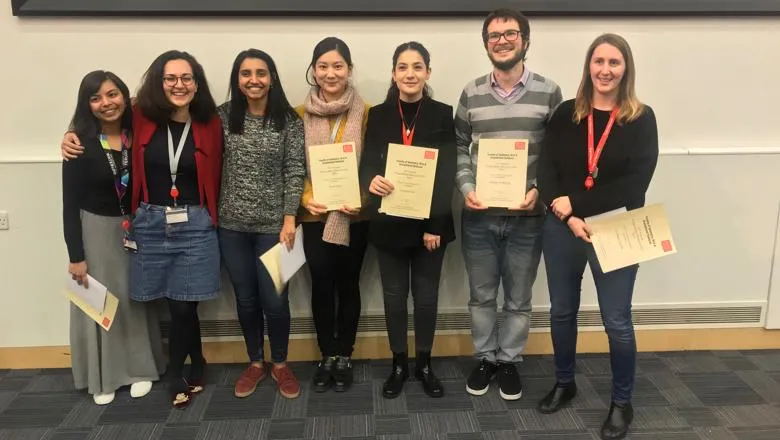
The 21st annual Postgraduate Research Day was held at the Faculty of Dentistry, Oral & Craniofacial Sciences on Wednesday 13 March. Featuring new and exciting research from our second and third year students, this day is the one occasion a year when student research in the faculty is showcased.
A great opportunity to share the exciting and innovative research that is being undertaken by students and staff, the day included presentations from a broad range of PGR students on their work. Building on the success of last year, one of the key criteria for presentations this year was accessibility and students were challenged to present their research to a mixed expert/non-expert audience.
Dr Sasha Scambler, Senior Lecturer in Sociology and co-organiser of the event said: “Year on year the quality of the presentations has improved and this year was no exception with presentations showcasing both the breadth and depth of the research carried out in the Faculty, and the communication skills of our students. As always the day would not have happened without the support of all of the volunteer judges and chairs as well as those presenting, so a huge thank you to all of those involved for an interesting and informative day.”
Professor Agi Grigoriadis, Associate Dean for Postgraduate Research said: “I just wanted to thank all our PGR students and extend my own congratulations to you for making this year’s PGR day so successful. You all made a huge effort and the talks were of especially high standard. The event keeps getting better every year, so well done. I hope all involved also found it equally enjoyable and rewarding. Keep up the great work.”
“Huge thanks also goes to Dr Sasha Scambler for organising another wonderful day and putting together an excellent programme.”
Congratulations to the students who won each session (full details below).
The winners from the sessions were:
Session 1:
Ruth Moon
Chromodomain helicase DNA-binding (Chd) proteins and inner ear development
Centre for Craniofacial and Regenerative Biology
Prof Andrea Streit, Prof Karen Steel
Members of the chromodomain-helicase (Chd) protein family are among the major ATP-dependent, chromatin remodeling factors. Mutations in several Chds are associated with human diseases that affect neural development and hearing. However, the exact mechanism by which they function in the developing inner ear is unknown. This study systematically analyses the function of Chd family members in the inner ear to understand the potential causes of hearing loss in mouse and thus provide new insight into hearing defects in patients.
The first aim of this study was to characterize the cell type specific expression of different Chd family members in the developing ear. While Chd7 is expressed throughout ear development (Hurd et al., 2010), Chd3 and Chd4 proteins are absent in the prosensory epithelium, but are later dynamically expressed in developing hair cells, various supporting cells and SGNs in partially overlapping patterns.
To establish the role of Chd7 in spiral ganglion neurons (SGNs), which innervate the sensory hair cells in the ear and project to the auditory nuclei in the brain, I used conditional Chd7 deletion in mouse SGNs. Loss of Chd7 does not affect early postnatal development, but during the first postnatal week SGNs begin to decline as compared to controls. In addition, many SGN axons show mistargeting and pathfinding defects. Together, this project may provide new insight into the mechanisms controlling ear development and those underlying hearing loss.
Session 2:
Fereshteh Sari
Molecular control of inner ear development: downstream of the SIX1 transcription factor
Centre for Craniofacial and Regenerative Biology
Professor Andrea Streit, Dr. Cynthia Andoniadou
The ear is responsible for hearing and balance, and the sensory cells that capture this information from the environment are located in the inner ear. During development, the entire inner ear originates from a simple epithelium in the ectoderm next to the hindbrain, the otic placode. The transcription factor SIX1 and its co-factor EYA1 are strongly expressed in the otic placode and play an important role in ear formation. However, the exact mechanisms downstream of these factors remain to be established. This project focuses on the identification and characterisation of SIX1 targets to provide insight into the genetic network controlled by the SIX1/EYA1 complex during inner ear development. We find that SIX1 putative targets are highly enriched in ear progenitors, and Six1 gain- and loss-of-function experiments in chick and frog reveal that they are indeed regulated by Six1. Characterisation of the regulatory regions that drive target gene expression in the ear shows that their in vivo activity depends on Six1 input. It is therefore likely that these genes are direct SIX1 targets and mediate its function during ear development.
Session 3:
Geraldine Jowett
Unraveling the impact of Th1 innate lymphoid cells on the intestinal stem cell crypt and its peri-cellular environment
Centre for Craniofacial and Regenerative Biology
Eileen Gentleman and Joana Neves
Inflammatory bowel disease (IBD) is a multifactorial disease that arises when the delicate homeostatic balance between the microbiome, immune system, and intestinal epithelial barrier integrity is disrupted by environmental factors or genetic predisposition. Anti-inflammatory biologics are the predominant form of treatment, yet they lack long term efficacy and penetrance, working in only a third of patients. This project proposes a reductionist, in vitro model of the gut-immune-environment to study the aetiology of IBD, proposing that new therapeutic strategies targetting epithelial integrity and extracellular matrix remodelling may be promising new approaches to treat this devastating disease.
Mouse gut organoids were co-cultured with primary intestinal type1 innate lymphoid cells (ILC1), as this novel, mucosal-tissue-resident sentinel cell type is enriched in inflamed gut biopsies from Crohn’s Disease patients, yet the cause and impact of this accumulation still requires investigation. RNA-sequencing of epithelial cells after co-culture with ILC1 in homeostatic conditions revealed dramatic changes in intestinal stem cell niche phenotype, e.g. in CD44 variant expression. Additionally, the extracellular matrix (Matrigel) in these cultures was fully degraded. The increased organoid growth and survival runs counter to the anticipated cell-death predicted by IFNy, the cytokine associated with ILC1. Thus, the next steps for this project are to elucidate novel mechanisms of interaction between ILC1 and epithelial cells predicted by this dataset, and to use a fully synthetic, inducably MMP-degradable or non-degradable PEG-hydrogel to tease apart the role that extracellular matrix remodeling plays in inducing the observed changes to the intestinal stem cell crypt.
Session 4:
Rupali Lav
Role of Wnt Pathway in tooth root development
Centre for Craniofacial and Regenerative Biology
Professor Abigail S Tucker and Professor Paul Sharpe
Although the initial stages of tooth development are well studied, little is known about root formation, specially the apex which forms a crucial channel through which blood vessels and nerves enter the vital dental pulp. It has been shown that a localized population of stem cells situated apical to the developing root (apical papilla) plays a crucial role in the formation of the dental root. The Wnt signaling pathway plays an important role in the specification of stem cells during initial stages of tooth organogenesis, however, its influence on the stem cells of the apical papilla remains unknown. This investigation focuses on localizing putative stem/progenitor cells in the developing murine tooth root. Here we have followed a developmental time course of murine root development, concentrating on the stages when the root apex develops, and have investigated how putative stem cell populations are established.
Session 5:
Nadhrah Ali
Stapes Development and Otosclerosis
Centre for Craniofacial and Regenerative Biology
Professor Abigail Tucker & Dr. Dan Jiang
The stapes is the smallest bone in the body, responsible for transferring sounds from the outer ear to the inner ear. The wide range of sounds transmitted is due to the stapes being able to vibrate freely in many directions. Otosclerosis is a disease where excess bone is deposited in the middle ear cavity. When the surplus bone grows on or around the stapes, the motion of the stapes is hindered, resulting in hearing loss. Although otosclerosis can be managed by surgery, little is known about the disease itself.
By understanding how the stapes develops normally, and which key genes are involved, this project aims to link development with possible mechanisms by which otosclerosis occurs. This has used the mouse as a model for development, with both wild-type and transgenic lines employed to answer questions about the ossification and remodeling of the stapes, as well as the role of genes involved in otosclerosis during development.
Session 6:
Mona Mozaffari
Understanding Canal Atresia: Early Development of the Mammalian Ear Canal
Centre for Craniofacial and Regenerative Biology
Abigail Tucker, Dan Jiang
Defects in ear canal development can cause severe hearing loss, however very little is known about how the canal initiates, extends and opens. In this study we show that the ear canal undergoes a complex system of closure and reopening as it forms. The more superficial part of the canal forms from an open primary canal, initiating at the junction between the first and second arch, which later collapsed and then reopened. In contrast, the deeper part of the canal formed from a solid meatal plate that extended from the primary canal into the first arch and later opened. As the ear canal developed, the different parts displayed distinct patterns of proliferation and keratin expression, with collapse of the primary canal linked to loss of periderm. When the primary canal was cultured at the timepoint of closure, pharmaceutical inhibition of apoptosis led to persistence of periderm and a failure to close. Likewise, in GHL3 -/- mice with deficient periderm an early closure of the primary canal was observed.
Final opening of the canal was triggered by terminal differentiation of the epithelium. Interestingly, the meatal plate opened asymmetrically, associated with differential proliferation, to create the thin outer surface of the ear-drum. Understanding these complex processes involved in canal development can shed light on the underlying causes of canal atresia.
Session 7:
Davide A Martella
NANOBIOPSY: Molecular Classification of Tumours by Spectroscopic Analysis of Tissue Replicas on Nanoneedles
Centre for Craniofacial and Regenerative Biology
Dr. Ciro Chiappini, Prof. David Richards, Dr. Mads Bergholt
Precision oncology aims to predict the optimal therapy for each patient, by tailoring treatments to the individual characteristics of each malignancy. Some key resources to tackle this challenge are genomics and molecular diagnostics. Molecular diagnostics evaluates the genome, transcriptome, proteome or metabolome of diseased tissue as discriminants of the tumour class. Yet, it largely relies on biomarker detection, which requires labour-intensive protocols and provides limited information on the overall molecular signature of tumours and their heterogeneity.
These restrictions negatively affect its diagnostic efficacy, by limiting sampling frequency and area, along with the number of observable biomarkers per sample.
NANOBIOPSY develops a platform for accurate, minimally invasive, label-free mapping of the molecular profile of tumour. This map allows stratifying tumours based on their molecular composition and spatial distribution (heterogeneity). It helps in cases such as primary malignant brain tumours, in which the current restrictions in sampling and number of considered biomarkers hinder the use of molecular diagnostics for preoperative and postoperative evaluation, as well as for surgical guidance.
In our approach, 8x8mm chips patterned with ordered arrays of porous silicon nanoneedles (nNs) are pressed onto the tissue to generate a replica of its molecular composition. This approach reduces the need for biopsies and significantly reduces invasiveness. The replica is analysed by a combination of Raman spectroscopy and mass spectrometry imaging, to combine high spatial and mass resolution. The stratification of clinical samples according to the shared molecular profile is performed through machine learning algorithms and the results are compared with histology.