What is Cardiac Resynchronisation therapy?
Cardiac resynchronisation therapy is a common procedure offered to patients who suffer from heart failure for which drug management alone is not effective. The condition prevents the heart from beating regularly due to uncoordinated electrical activity. This means the organ struggles to circulate blood and oxygen around the body, resulting in patients straining with simple, everyday tasks.
The aim of the therapy is to resynchronize the contractions of the heart ventricles by implanting a device in the patient. Three pacing leads must be placed into the right atrium, the right ventricle, and the left ventricle of the patient’s heart which can deliver electrical signals to the heart muscle.
Lead placement for the left ventricle specifically has a critical influence on the procedure, and sub-optimal placement can cause poor outcomes in around 40% of cases. However, the optimal location is highly patient specific and can be affected by both anatomy and disease factors such as the presence of scar tissue.
How can software be used for patient benefit?
The researchers have developed an integrated guidance system which fuses pre-operative imaging data in real time during the insertion of the leads, typically performed using a catheter under X-Ray fluoroscopy guidance.
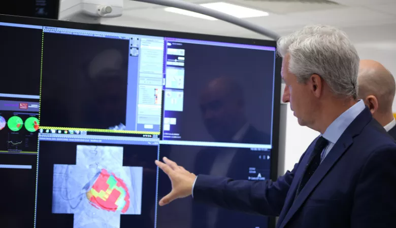
The software creates a patient specific left ventricle model using MRI scan data which takes into account critical metrics including the presence of scar tissue and the latest point of mechanical activity. Automatic segmentation capabilities have also been incorporated into the software through machine learning techniques.
This allows surgeons to rapidly calculate the optimal position for the lead placement and use this as guidance during therapy by fusing the information with the fluoroscopic images.
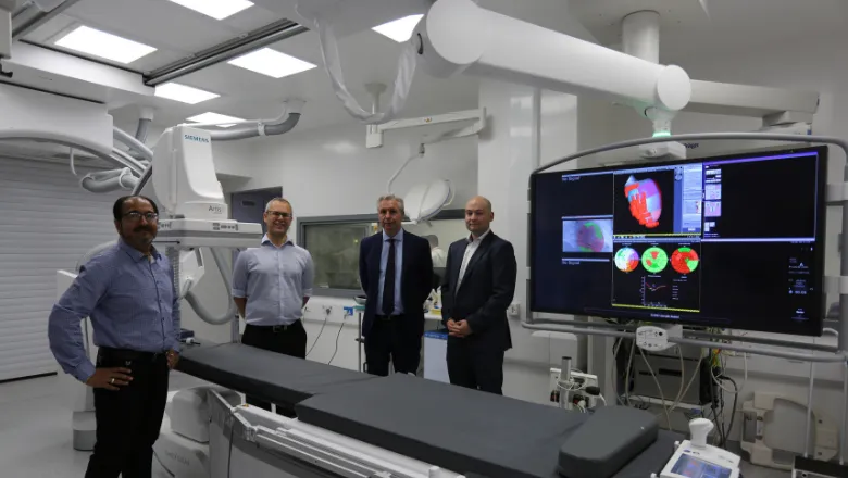
The system has been developed and tested using an XMR facility, a suite which combines both an MRI scanner and a catheter lab in adjoining rooms. Patients are scanned in the MRI directly before their procedure and the software is able to process the pre-operative data by the time it takes to transfer the patient to the catheter lab, demonstrating the significant time-saving possibilities of this system.
A new image guided system
During this project the researchers were able to demonstrate proof of concept for 20 patients. Whilst exciting, wider scale trials are required to fully demonstrate the feasibility and efficacy of this integrated guidance platform. However, as the system has been designed to fit within the current clinical workflow any further translation of this research should be widely suitable for other teams and treatment centres to use. With cardiovascular disease being a global condition, the potential for creating impact and improving quality of life for those with heart failure is vast.
Concepts and information presented are based on research and are not commercially available. Due to regulatory reasons, the future availability cannot be guaranteed.
Awards
Information Processing in Computer-Assisted Interventions: Bench to Bedside Award
European Heart Rhythm Association: Inventors awards (runner up)
Statistical Atlases and Computational Modeling of the Heart: Best Paper Award
Relevant papers:
- Real-Time X-MRI-Guided Left Ventricular Lead Implantation for Targeted Delivery of Cardiac Resynchronization Therapy.BeharJM, Mountney P, Toth D, Reiml S, Panayiotou M, Brost A, Fahn B, Karim R, Claridge S, Jackson T, Sieniewicz B, Patel N, O’Neill M, Razavi R, Rhode K, RinaldiJACC Clin Electrophysiol. 2017 Aug;3(8):803-814. doi: 10.1016/j.jacep.2017.01.018. Epub 2017 Apr 26.
- A Planning and Guidance Platform for Cardiac Resynchronization Therapy.Mountney P,Behar JM, Toth D, Panayiotou M, Reiml S, Jolly MP, Karim R, Zhang L, Brost A, Rinaldi CA, Rhode K.IEEE Trans Med Imaging. 2017 Nov;36(11):2366-2375. doi: 10.1109/TMI.2017.2720158. Epub 2017 Jun 27.
- 3D/2D Registration with superabundant vessel reconstruction for cardiac resynchronization therapy.Toth D, Panayiotou M, Brost A,Behar JM, Rinaldi CA, Rhode KS, Mountney P.Med Image Anal. 2017 Dec;42:160-172. doi: 10.1016/j.media.2017.08.001. Epub 2017 Aug 5.
- 3D/2D model-to-image registration by imitation learning for cardiac procedures.Toth, D., Miao, S., Kurzendorfer, T., Rinaldi, C.A., Liao, R., Mansi, T., Rhode, K. and Mountney, P., 2018.. International journal of computer assisted radiology and surgery, pp.1-9.