16 December 2019
Biomedical Engineering & Imaging Sciences researchers developing hyperspectral imaging camera
Lightweight hyperspectral imaging cameras could offer surgeons real-time tissue differentiation
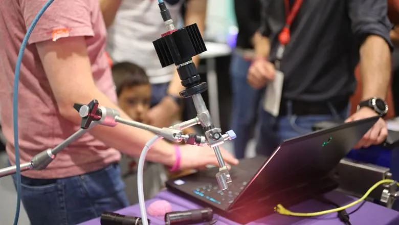
Researchers at the School of Biomedical Engineering & Imaging Sciences, in collaboration with University College London, are for the first time developing a novel light-weight camera device that is easily integrated into a clinical workflow to provide real-time tissue differentiation between tumour tissue and normal tissue helping make operations safer and more effective.
It is often very difficult for clinicians to differentiate between tumours and surrounding critical structures such as nerves and blood vessels.
Some tumours sit outside the brain and push on it, displacing normal structures. The challenge is to take out as much of that tumour as can be done safely without disrupting or damaging the surrounding structures.
A hyperspectral imaging system would help to differentiate between critical structures and tumour, while also providing the surgeon with real-time information.
“The advantage of this system is that it could differentiate tissues within a large field of view and the computer-generated tissue labels could then be displayed as an image overlay on top of the surgeon’s regular image,” lead researcher Mr Jonathan Shapey, said.
Frequently, surgeons make decisions based on what they can see with the naked eye, often using a microscope which allows them to see a tumour in high definition. But there are of course inherent limitations with this technique.
Within the optical spectrum there is a visible band between 400-600 nanometres – the spectrum of what we perceive as blue, green and red light. Beyond 600 nanometres there is infra-red information which our eyes are not receptive to.
However, such ‘invisible’ bands contain valuable information that could be exploited to aid clinical decision making.
Cameras like the hyperspectral imaging cameras acquire many more bands than just three in the visible spectrum, i.e. red, blue and green, but also in those areas we do not have the capacity to see.
Postdoctoral research associate Dr Yijing Xie analyses the optical property of tissues to help determine which camera should be used and what hardware could be used for this application.
Without knowing the optical properties of tissues, researchers are unable to identify which camera or which light source can be used – the camera needs to be tailored to meet each application.
“The aim is to classify the structures seen within the surgical area but to do this we need to map what it is that makes a tumour tissue appear as tumour tissue and importantly, how this differs from a healthy tissue type,” Dr Xie said.
The hyperspectral imaging cameras can also open the door to new means of measuring established characteristics such as oxygenation levels which can be extracted from the infra-red bands.
Postdoctoral researcher Dr Michael Ebner, who works on the computational aspect of the research, said that the new methods will allow for real-time visualization of tissue boundary and tissue property information (such as oxygenation saturation levels) which is currently not available to the surgeon.
“This makes the decision-making process faster and safer because you have more information right in front of you and it helps to react in case something unexpected happens,” Dr Ebner said.
“We also hope that with more information, that is instead of three bands we use 25, 100 plus bands, we can really do much more reliable tissue differentiation that can be done currently in clinical practice.”
Research Associate Dr Eli Nabavi, who designed and developed the hyperspectral imaging system, said the cameras can be easily integrated into the clinical workflow.
“The clinical and engineering teams work closely together as a multidisciplinary team to ensure that the device integrates well into the existing clinical and surgical workflow,” Dr Navabi said.
For the future, the team will continue to work on developing the system’s ability to better understand tissues’ optical properties.
Mr Shapey said: “This novel and lightweight hyperspectral imaging camera system presents a tremendous opportunity to clinicians by equipping them with a tool that will ensure more accurate tumour identification and safer surgery.”