She's doted on by everyone and she's just thriving – and it's all down to these specialists and this technology. It's amazing what they do, it's lifesaving.
Kirbi-Lea Pettitt, mother of Violet-Vienna
25 July 2019
Detecting foetal heart defects
A pioneering method developed by King’s researchers and clinicians helps save babies’ lives.

When a heart problem is suspected in a baby before birth, the mother will be referred to a foetal cardiologist for a detailed ultrasound of their child’s heart. In some cases, the details of these conditions can be difficult to diagnose with ultrasound, and to date there have been no other reliable alternatives.
This means the family may have to wait until their baby has been delivered to get more information about their heart condition and what kind of treatment they will need.
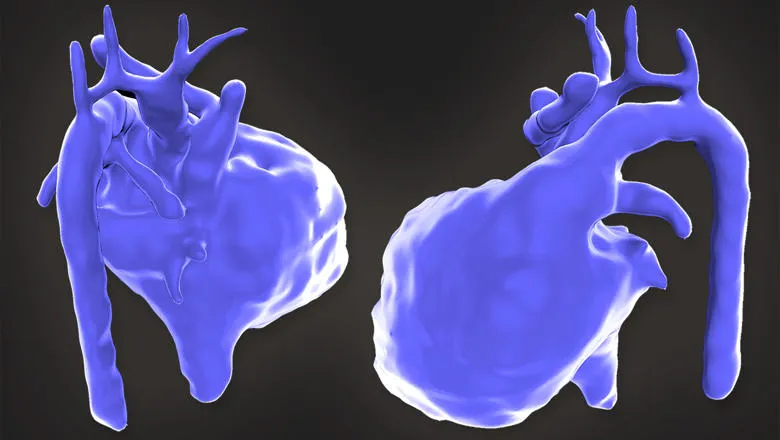
Our research team at the School of Biomedical Engineering & Imaging Sciences, along with clinicians at the Evelina London Children’s Hospital, have developed new techniques using MRI scans performed in pregnancy to produce high-resolution 3D images of babies’ hearts in the womb.
These detailed images, along with ultrasound, allow foetal cardiologists to get a more accurate understanding of a baby’s heart and plan for the right treatment after they are born.
One such baby is Violet-Vienna, who developed abnormalities in the blood vessels around her heart while she was still inside her mother’s womb. Problems were initially detected when her mother, Kirbi-Lea Pettitt, went for a routine ultrasound scan 20 weeks into her pregnancy. Kirby-Lea took part in a study that allowed doctors to look at her baby’s heart in vivid detail and plan how to save Violet-Vienna's life after she arrived in the world.
It is hoped that this type of technology will lead to better outcomes for children and families affected by congenital heart disease.
Learn more about this pioneering method and how the King’s community is serving society in the university’s Service annual report 2018-19.
