22 March 2019
Fetal heart abnormalities detailed before birth by new 3D images
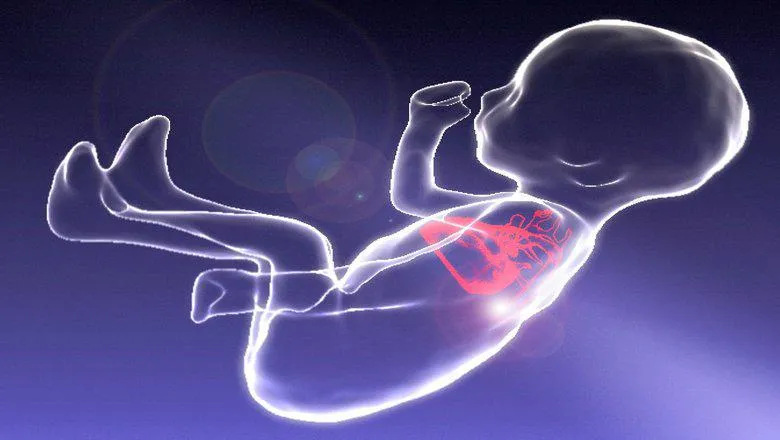
Scientists from King’s College London and the Evelina London Children’s Hospital at Guy’s and St Thomas’ NHS Trust have developed a new method using MRI for producing detailed 3D images of the fetal heart, to improve the diagnosis of congenital heart disease before birth.
When congenital heart disease is suspected in a baby before they are born, clinicians using ultrasound are not always able to diagnose all details of the condition due to the technical challenges involved with imaging the tiny, fast-moving baby. This is important when an abnormality involves the blood vessels around the heart.
In the paper published today in The Lancet, the team successfully used a new computer processing method to make the standard MRI images that are often unclear because of the moving baby into clear three-dimensional models.
The 3D models applied to 85 pregnant women provided clinicians with reliable, high-resolution information about the fetal heart, helping to optimise treatment for the affected babies after birth.
First author, Dr David Lloyd, Clinical Research Fellow at King’s said: “Our hope is that this approach will now become standard practice for the Evelina fetal cardiology team who make a prenatal diagnosis in 400 babies each year. This will also improve the care of over 150 babies each year who deliver at St Thomas Hospital with known congenital heart disease.”
John Simpson, Professor of Paediatric and Fetal Cardiology at Evelina London, said: "Three dimensional MRI revolutionise the type of information we can obtain before babies are born. This impacts directly on care we provide after birth and provides new insights into structural heart defects before birth."
Senior author, Professor Reza Razavi, Professor of Paediatric Cardiovascular Science and Consultant Paediatric Cardiologist, said: “Application of this novel computing technology has enabled for the first time MRI scanning to really help with clarifying the diagnosis in a subgroup of babies, particularly when the vessels around the heart are involved.”
The team is now working to combine this 3D imaging with other advanced ultrasound and MRI techniques, to try to understand why some babies go on to develop more severe forms of congenital heart disease than others.
The full paper is available online.
In this story

Vice President (Research)