13 March 2019
Growth and mineralogy in dental plates of the holocephalan Harriotta raleighana (Chondrichthyes)
Novel dentine and conserved patterning combine to create a unique chondrichthyan dentition
.x164ec2e4.jpg?w=780&h=1170&crop=780,440,0,365&f=webp)
We describe a new type of extra hard, hypermineralised dentine, exposed at the oral surface that is made to order from specialized odontoblasts, deep within the dentine of all six compound dental plates. All are entirely composed of dentine and all are constantly renewed at the aboral surface, first the trabecular dentine makes spaces in a preformed pattern. These spaces are only subsequently filled with the hypermineralised dentine, mineralizing late and suddenly relative to the ordinary dentine. This entire group of cartilaginous fish have lost the developmental mechanism to make separate, replaceable teeth that is a feature defining the dentitions of all sharks and rays. Our research describes how the Spookshark, HarriottarRaleighana, a benthic feeder in the deep ocean, manages without teeth, and hence without the very hard coronal enameloid. Instead they make this unique form of dentine in a pattern that is shown at the oral surface as ordered projections that are resistant to wear (Figs, 1, 2, 3). Many of these features are novel, presenting a unique dentition among chondrichthyans, including new modes of dentine deposition and mineralization that may prove to be general for holocephalans more broadly. We could ask, “How do these fish manage without teeth”?
A key to understanding how this statodont dentition (non-shedding, with replacement growth from within) functions without teeth is the arrangement of the hypermineralized dentine. There is a pattern and shape to separate regions of this dentine called ovoids and tritors that are encased within a less mineralized, trabecular dentine. These ovoids and tritors are preformed as collagenous capsules, with mineral empty spaces (Fig.3), all are supported within the trabecular dentine framework of the dental plates. Associated with the layer of specialized odontobast cells making this hypermineralized dentine is a notable vascular system, especially for the tritors, it comprises tubes of vascular pulpal tissues, and as suggested, localized stem cells. This pattern is reflected in the occlusal wear surface and ensures an oral surface of projecting hard elements, within less hard dentine, occlusally arranged as adapted for feeding (Fig. 4).
Quantitative elemental plots through a series of developing ovoids showed elevated Mg readings relative to the other elements in the least mineralized parts of the dental plate and likely represents Mg-rich and poorly crystalline hydroxyapatite. This mineral forms within a dominant array of cell tubules, where organic matrix is scarce, with this massive array of tubules emanating from the specialized odontoblast cell bodies that line the surfaces making the hypermineralized dentine. This elemental composition correlates very early on in the mineralization sequence with a non-crystalline phase of optically dense, non-birefringent, minimal mineral density material, in the early mineralizing ovoids (Fig. 3 D, E).
We show that this specialized dentine is unique to dental hard tissues in its composition as a magnesium rich tricalcium phoshate (MTCP, ß-Ca3 Mgx(PO4)2), similar to a mineral called Whitlockite. Also novel is the developmental mineralization process, initially formed as Mg-rich oxide in amorphous phase (Fig. 3D) directly transforming into irregular crystalline granules, then progressively replaced by Mg Whitlockite. The proposed name for this mineralized tissue is ‘whitlockin’, providing a dentition that is unique for the whole group of holocephalans, as a second holocephalan, Chimera, is known to have similar HD that contains whitlockite, amongst trabecular dentine dominated by hydroxyapatite. The larger, specialized cells (whitloblasts) that secrete the whitlockin provide multitudes of cell processes that penetrate deep into the early mineralizing tissue. These tubules from whitloblasts give rise to membranous saccules and vesicles that must be the key to such a novel mode for mineralizing the specialized dentine as whitlockin.
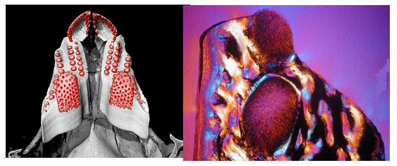
Figure 1. Upper jaw of Harriotta raleighana with four dental pates in situ, mCT scanned and rendered for density differences, most mineral dense segmented as red, shows in the caudal plate, rows of ovoids rostral to the tritor seen as a larger mass of tissue with a punctate surface, where large vascular canals intrinsic to this tissue are filled in with less dense, circumvascular dentine. The paired rostral plates contain only many, rows of ovoids, no tritoral mass of hypermineralized dentine supplied with many, organised, vascular canals, representing the dental pulp.
Figure 2. Section through the surface to illustrate two of the ovoids, one exposed at oral surface surface. All are supported by trabecular centine, which is birefringent in polarised light, using a gypsum plate that shows in blue and yellow the opposite orientation of collagen fibres as the basis of this dentine, red is neutral. This hypermineralized dentine has no fibrous matrix, is packed with odontoblast tubules, and surrounded by a capsule of circular fibres. The mineral crystals are not oriented and are composed of a Magnesium rich, calcium phosphate, similar to Whitlockite, and unlike the predominant mineral of the dentine as hydroxyapatite.

Fig. 3A-E, are of section views through one dental plate, with three ways of viewing the hardest tissues of the dentine, ovoids in all and tritor in C, which is a virtual section from a CT scanned, 3D rendered lower jaw dental plate. A, B are taken of the surface in reflected light, of the whole sectioned dental plate in the jaw cartilage and a high power view of the upper set of ovoids. Organization is a stack of ovoids renewed from those forming at the aboral surface and increasing in translucency that visualises the late hypermineralization of them towards the worn surface. In C the high mineral density is seen in both ovoids and tritor, relative to the empty aboral ovoids in the stack, all surrounded by lower density trabecular dentine. D, E, are optical micrographs through an upper jaw dental plate, of the same two ovoids, with crossed polars (see Fig.2). In D, the more oral ovoid has transitioned to highly mineralized with some oriented birefringence (E), relatively the lower ovoid is non-birefringent and opaque, (D) black region, and retains the Mg rich amorphous phase that turns into granular crystal in the process of hypermineralization. In A, B, the worn oral surface also shows the third tissue type, we term sclerotic dentine (See Fig.4, yellow) as the trabecular dentine has become compact and with increased mineral. In this way the dental plates compensate, and adapt to function, without any teeth.
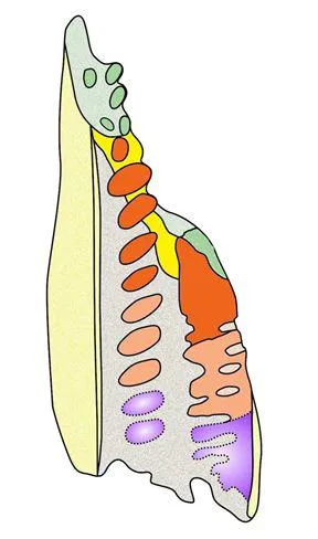
Figure 4 – Diagram of tissue distribution, both ovoids and tritor, represents how they are renewed at the aboral surface and become increasingly mineralized. Colours represent these stages of growth forming hypermineralized dentine (HD green) at the worn oral surface and orange in section view. This HD tissue (purple) at the aboral surface is pre-formed for renewal as a pattern in the trabecular dentine (grey), then it transitions to high mineral content, low organic (pale orange), then hypermineralized dentine (orange) formed my masses of ramifying, odontoblast tubules from specialized cells at the secretory surfaces. A third tissue is sclerotic dentine (yellow), formed near the worn surface related to wear (see Fig. 3 A, B).
In this story

Professor of Evolutionary Dentoskeletal Biology