
Bringing the Human to the Artificial
Explore artificial intelligence and automated decision-making at this King's exhibition.
28 April 2023
Read about King's work on how AI is being used in oral micro-vascular diagnostic imaging, as featured in the Bringing the Human to the Artificial exhibition.
In healthy mouths, the smallest blood vessels (capillaries) form tiny loops just under the mucosal surface. This allows the surface lining to survive. In disease, however, these loops change. We are using AI to observe capillaries and to identify different tissue patterns without human intervention and bias.
In inflammation, capillaries become wider and more can open up, which is why inflamed mucosa looks red; in cancer conditions, the micro-vessel shapes change further and new vessels are driven to grow (angiogenesis) by the effect of the cancer itself as it demands constant energy supplies to grow.
Over the last decades as medical instruments have been developed to examine tissues in vivo and in situ, several qualitative reporting and descriptive classification schemes have been introduced. However, these are fraught with human observer bias and potential inconsistency of interpretation as different opinion enters the classification assessment.
We have developed imaging instruments that can see the capillary circulation directly. Using machine learning, we are starting to be able to segregate malignant, pre-malignant, inflammatory and normal tissue patterns, without human intervention or the biases of human image assessment. This should allow us to ultimately look for indications of malignancy, without any local anaesthetic or other surgical tissue harvesting being necessary.
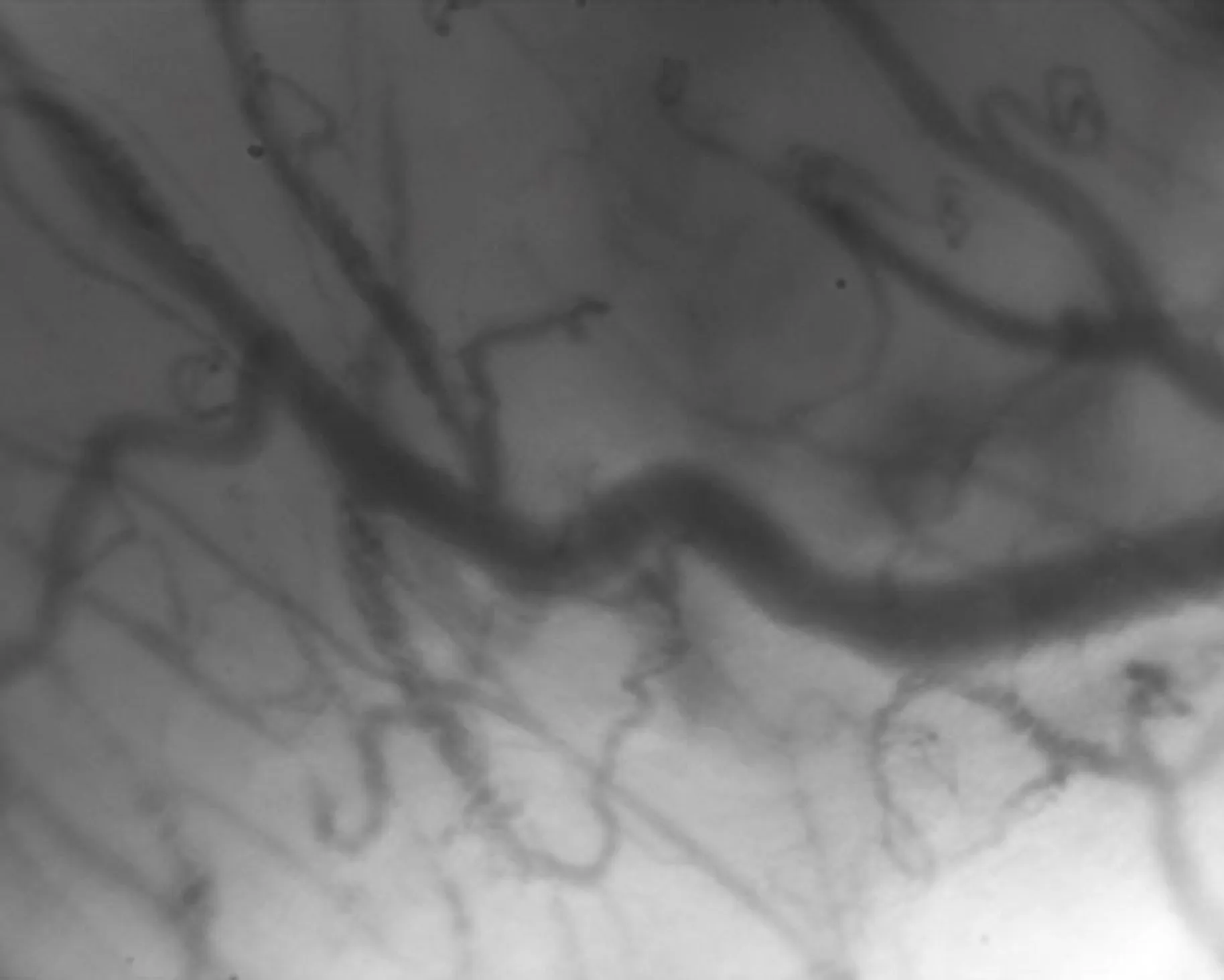
Example of how the raw image (A) is processed to a clean image and the software plots the map (B), then the map is abstracted (C), and the data characteristics are extracted from the map, ready for AI interpretation of characteristics of the image (D).
A: initial raw microvascular capillary image. B: modelling and analysis of the skeleton (derived from binarising image A after background subtraction), superimposed to optimised version of image A. C: model of the microvascularisation composed of different vectoral elements. D: amplified image from C with Jnc (junction), Ext (extremity), Seg (segment), IsE (isolated element), Br (branch) and lp (loop). Vectorial elements are indicated as some examples of the quantitative summary characteristics extracted from the raw image in A, that can be analysed objectively in the process, to indicate the likely pathology driving the behaviour of the micro-circulation.
Richard Cook, Tim Watson
Centre for Oral, Clinical & Translational Sciences, Faculty of Dentistry, Oral & Craniofacial Sciences, King’s College London
Pedro Bastos
Centre for Oral, Clinical & Translational Sciences, Faculty of Dentistry, Oral & Craniofacial Sciences, King’s College London and Homerton Hospital Healthcare Trust
Gilles Carpentier
Université Paris-Est
King’s College London, Medical Research Council, London Development Agency, Guy’s Charity, Oral Dental Research Trust
Guy’s and St Thomas’ NHS Foundation Trust Dental Hospital
Visit Richard Cook's King's profile
Visit Tim Watson's King's profile
Read Real-time optical vascular imaging: a method to assess the microvascular circulation of myofascial free flaps used in the head and neck region in the International Journal of Oral & Maxillofacial Surgery

Professor in Diagnostic technologies / Hon Consultant in Oral Medicine

Professor of Biomaterials & Restorative Dentistry
Explore artificial intelligence and automated decision-making at this King's exhibition.