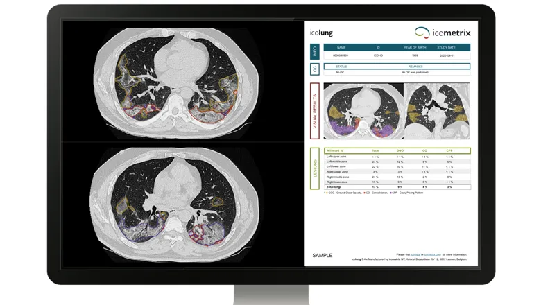
The size of lesions detected and their localisation can help in the management of COVID-19 patients, a key benefit of using CT scans along with the AI system for more patients.
Lucas Fidon, PhD student
13 May 2020
Icolung, an imaging AI tool for chest CT to support patient and resource management during COVID-19
PhD student from School of Biomedical Engineering & Imaging Sciences has algorithm integrated into AI software and is now available for clinical use in Europe and the US
In response to the COVID-19 pandemic, icometrix and academic partners, including King’s College London School of Biomedical Engineering & Imaging Sciences, developed icolung.
icolung is the first CE-marked AI solution for quantifying lung pathology on chest CT scans in COVID-19 patients resulting from a COVID-19 collaboration.
The solution can assist in the risk-based triage of COVID-19 patients by quantifying the degree of lung involvement.
The degree of lung involvement on CT is an essential aspect of the pulmonary status as it correlates with the severity of the disease and the outcome (Colombi et al. 2020; Revel et al. 2020).
For this reason, CT provides insights into the pulmonary status of COVID-19 patients with moderate to severe symptoms and can facilitate the risk stratification.
Chest CT scans allow identification of the location and extent of different types of lesions in the lungs.
But so far, available AI systems for chest CT scans were only able to differentiate lesions from normal pulmonary tissue, without distinguishing different lesion types.
icolung provides an objective quantification of the degree of lung involvement on CT in COVID-19 patients, in the different lung regions and per lesion type.
School of Biomedical Engineering & Imaging Sciences Ph.D. student, Lucas Fidon, who is completing a research secondment at icometrix, is developing part of the AI-based software which automatically delineates lung lesions in COVID-19 chest scans, a task that would take even experienced radiologists hours to achieve.
Quantification of the lung involvement in COVID-19 patients is found to increase the specificity to predict ICU admission or death, meaning that clinicians will be able to better identify patients who can be sent home or to a non-ICU bed (Colombi et al. 2020).
Pulmonologists, emergency physicians, and intensive care physicians could, therefore, make a better-informed decision when knowing the extent of pulmonary infiltration in COVID-19 patients.
“AI allows obtaining a 3D delineation of the different lesions in minutes and with higher accuracy,” Mr Fidon, said.
References
- Colombi D, Bodini FC, Petrini M, Maffi G, et al Well-aerated Lung on Admitting Chest CT to Predict Adverse Outcome in COVID-19 Pneumonia. Radiology. 2020 Apr 17:201433. doi: 10.1148/radiol.2020201433.
- Revel, M., Parkar, A.P., Prosch, H. et al. COVID-19 patients and the radiology department – advice from the European Society of Radiology (ESR) and the European Society of Thoracic Imaging (ESTI). Eur Radiol (2020). https://doi.org/10.1007/s00330-020-06865-y