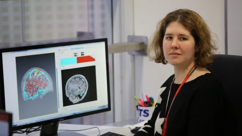
My work at King’s is trying to take EpiNav further and expand the concept of surgical navigation in neurosurgery for other diseases, not just epilepsy.
Rachel Sparks
05 February 2019
Interview with Rachel Sparks: Developing computer models for brain surgery
This month we interview Rachel Sparks, Lecturer in the Department of Surgical and Interventional Engineering at King's. Read on below to find out how her work on developing computer models from large data sets to help shape the future of surgery, and EpiNav, a tool that focuses on providing neurosurgeons clearer pathways in surgery.

Can you tell us a little more about your background?
All my training has been in the realm of biomedical engineering. I started as an undergraduate looking at high-intensity focused ultrasound for neurosurgery application. During my PhD I focused on image analysis, exploring questions like, “Can we identify the same body part in both a MR and CT scan?”
I also did quite a bit of work on cancer diagnosis (e.g. can an MR image of a prostate or breast identify if there is a tumour, and if so, can we say whether it’s cancerous or benign?) After I wrapped up my PhD, I moved to UCL where I started working on a project called Epilepsy Navigation, or EpiNav. EpiNav focuses on providing neurosurgeons tools to help them plan surgeries, specifically in the context of epilepsy surgery which has a very complicated clinical pathway.
A multidisciplinary team of neurologists, electrophysiologists and a neurosurgeon first try to determine where seizures are coming from, and secondly, try to determine if they can safely remove it. The team takes quite a few MR scans to assess the patient’s anatomy and then implant electrodes to record brain activity and determine exactly where the seizures are coming from. Using these two bits of information, the team analyses where in the brain they want to operate on. A lot of what EpiNav is doing it is providing surgeons tools to co-register images across different modalities and to definitively assess where the electrodes ended up, relative to other parts of the patient’s anatomy. Essentially, in EpiNav you can build a very complicated and detailed 3D model of the patient’s brain.
One of the other applications where this technology would be very useful is tumour resection, where it is less about identifying where seizures are coming from and more about being able to assess the margin of the tumour and define precisely where to resect without causing damage to functional areas of the brain.
What research are you currently working on at King’s?
Refining parts of the EpiNav platform, in which there are three active areas. The first is around modelling tool biomechanics. The brain as an object moves and has some properties to it, and when you're inserting tools like electrodes or biopsy needles into the brain, two things can happen: the tool itself can move, or the tool can cause the brain to deform. We’re trying to build a biomechanical simulation so surgeons can see how the brain might deform based on the treatment that they’re planning.
The second area is much more around classic image analysis. A lot of what we're looking at is how can we develop methods for taking diffusion MRI and functional MRI and identifying parts of the brain that are important for cognition and motor function. This is useful because now surgeons have a sense of where not to go. A lot of this work is focused on how we can analyse these images in a consistent way so that regardless of which surgeon is using these images, they have a consistent answer of which part of the brain is functional. The second bit in this research is determining the best image to guarantee consistent results.
The third area we look at, which is very much focused on epilepsy, is trying to understand video-telemetry data. This is essentially placing electrodes in the patient’s brain that will allow their brain activity to be recorded, while also being video monitored. This data will be used to figure out where in the brain the seizure is coming from. We’re trying to develop tools to automate this analysis because at the moment patients are manually record over the course of one to two weeks – 50% of which the patient is sleeping, limiting surgeons to only focus on the areas in time where there might be seizures or when there's the most electrical activity. I am also working on the SEG analysis side, which is recording how electrical signals occur in the brain. Obviously, there's correlations between different brain regions at the same time and we’re trying to develop computational models to explain how electrical activity in the brain propagates to help us localise the start of the seizure.
Where do you think your research is headed in the next five years?
The thing I am most interested in is open craniotomy. A lot of what we’ve been working on is keyhole neurosurgery - drilling small holes in the brain and inserting small tools. These procedures are little bit easier to automate because the brain is less likely to deform. Open craniotomy is removing a larger piece of the skull. The brain behaves in more unpredictable ways, and it gets more complicated in terms of surgical planning, like brain shift. You can imagine that the brain acts like it’s sitting inside a water balloon. If you puncture the dura (the thin membrane surrounding the brain), it's like puncturing a water balloon. It will deflate and shift around because you’ve penetrated this thin membrane that helps keep all the cerebral spinal fluid close to the brain. Once that happens, all the pre-operative imaging you’ve taken to help identify where in the brain things are shifts relative to where the brain actually is. So, it’s much more complicated for neurosurgeons to understand where to go and where they can cut safely. While it is challenging, if we provide them tools where we can show where things are shifting, surgery can truly be improved. The overarching theme of my work is trying to develop software tools so surgeons know where to go and improve patient-outcomes in the long term because you can increase safety and advocacy at the same time.
What do you believe will be the next breakthrough in medical imaging?
Being able to develop computer models from large data sets that builds on several patient cases.
Surgeons are initially trained via an apprenticeship program, where they start their residency by getting paired with a more senior surgeon who trains them on a similar approach based on how they perform their own procedures. It’s really someone who has 20 years of experience trying to teach someone up and coming. It’s very hands on, but in some ways, it’s limited because the new generation of surgeons are being trained based on individual experience by the previous generation. Medicine is now bringing in more artificial intelligence and computing, not because the old methods are necessarily wrong, but because any individual surgeon has limited experience because of the individual cases only they have seen. So, being able to develop tools based on large data sets of several surgeries would provide more detailed and faster insight.
What advice would you give to other researchers in the engineering field, specifically women?
I think for anyone, but it can be especially hard for a woman, is being able to identify your expertise. Once you know that, find collaborators within the field who don’t have the same expertise as you, but who have compatible expertise. My expertise, for example, is developing software and computer models for representing data. I’ve had to develop collaborations with expert surgeons because I’m not a surgeon. I’m not an MR physicist, but I have expert MR physicists who do the image acquisition. To advance in the field, you need to have people who have skills you don’t have, but who you can collaborative on projects with. Especially as a woman, one of the things that can be hard is to be confident in what you know. Don’t be afraid to call yourself an expert in something if you’re very passionate about it.
