“The School’s outstanding research was very well represented at MICCAI this year and is reflective of a strong collaborative work ethic by the School’s researchers. Acceptance of submissions to MICCAI is extremely competitive, and I’m excited to have seen such a strong presence from the School this year.”
Professor Sebastien Ourselin
05 November 2019
King's School of Biomedical Engineering & Imaging Sciences has prominent representation at MICCAI 2019
A large cohort of students from the school attended MICCAI 2019
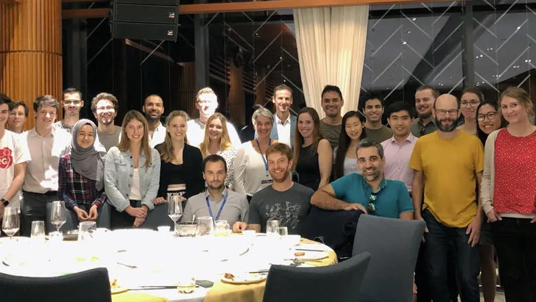
King’s School of Biomedical Engineering & Imaging Sciences was proudly represented by a cohort of more than 40 PhD and postdoctoral students who shared their research at this year’s MICCAI, the 22nd International Conference on Medical Image Computing and Computer Assisted Intervention which took place in Shenzhen, China, October 13th – 17th.
The annual MICCAI conference attracts world leading biomedical scientists, engineers, and clinicians from a wide range of disciplines associated with medical imaging and computer assisted intervention.
The conference series included three days of oral presentations, poster sessions and satellite events including workshops, tutorials and challenges as well as an industry forum on the days preceding and succeeding the conference.
Professor Julia Schnabel, Professor of Computational Imaging and Head of Research & Impact at King’s College London said the School’s impressive delegation of more than 40 researchers presented 17 papers at the main conference, including platform presentations from the iFIND and SmartHeart projects, with many more presentations at the satellite events.
Participants organised a number of MICCAI workshops and tutorials, including Computational Diffusion MRI, Computation and Visualisation, Domain Adaptation and Representation Transfer, Deep Learning, paediatric image analysis, statistical atlases , ultrasound imaging and machine learning.
Marc Modat, Senior Lecturer in Computational Imaging, said MICCAI was an opportunity to reinforce the position of the School by showing the exciting research happening.
“It also enabled people otherwise based in different locations to socialise during the conference as well as outside. Hopefully, this will make the links between the research groups/locations grow stronger,” he said.
King’s PhD student Lucas Fidon was part of the School participants who presented their poster.
Mr Fidon’s research looks at enforcing biological constraints in registration algorithms.
“The developed registration method was applied to the cardiac MRI and it turns out that it is interesting in a clinical workflow to track the myocardium,” Mr Fidon said.
“It is difficult to do this tracking with current methods because in the images there are no contrasts in the myocardium but we know that this muscle is incompressible, meaning that it can be deformed or warped but its volume will remain the same.”
Mr Fidon said there is no way that current algorithms fully satisfy this constraint, but his work seeks to identify ways to impose these constraints into the tracking algorithm.
His work proposes a new method in the algorithm that allowed a high accuracy in terms of incompressibility –several orders of magnitude better than previous methods.
PhD student Shu Wang also presented her poster on valve model making using 3D printing and its imaging applications.
Her poster described how to build ultrasound and MRI-compatible aortic valves compliant phantoms with a two-part mold technique using 3D printing.
“The simplicity of the manufacturing process and low cost of materials should enable an easy adoption of our proposed methodology,” Ms Wang said.
“Future research will focus on the extension of the method to cover a larger anatomical area, for example, aortic arch and the use of this phantom to validate the non-invasive assessment of blood pressure differences.”
Another success came by way of PhD student David Drobny who came second in the MICCAI CuRIOUS image registration challenge.
“This was the first time I attended MICCAI and it was a great experience starting off with winning a prize in a prestigious MICCAI challenge,” Mr Drobny said.
Mr Drobny used NiftyReg as software to compute the registration of 3D MRI and 3D ultrasound images in the context of brain surgery.
The submission to the challenge was first a paper that described the methods suggested to solve the challenge based on training data made available.
Test data were then published that were aligned with the methods described in the paper. Landmark coordinates after the registration were sent to the organisers who computed the distance to the reference landmarks. The average error of all landmarks per subject are used to generate a ranking of the teams.
Research Fellow Carole Sudre, whose work looks at vascular lesions when people age, was featured in the MICCAI Daily magazine prior to running the UNSURE workshop on the uncertainty for safe utilisation of machine learning in medical imaging.