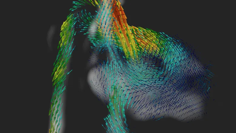
The results in the paper are exciting because no one has been able to look at the fetal heart using MRI in four dimensions like this. When we give these videos of the beating heart to the doctors, they are able to interact and examine the direction of blood flow instantly.
Dr Tom Roberts
05 October 2020
New method developed by researchers for in utero 4D blood flow visualization of baby hearts paves the way for better diagnosis of congenital heart disease
With further development, the method could become a new tool for aiding diagnosis of congenital heart disease where conventional methods like ultrasound might fail
Researchers at the School of Biomedical Engineering & Imaging Sciences have developed a new method for helping detect congenital heart disease of a baby in pregnant mothers using MRI. Existing in-utero approaches are compromised by fetal motion, but the novel method corrects the motion to present 4D visualizations of the heart depicting major vessels and blood flow circulation. With further development, the method could become a new tool for aiding diagnosis of congenital heart disease where conventional methods like ultrasound might fail.
For the first time, in a new paper published today in Nature Communications, researchers have been able to look at the fetal heart in four dimensions – the images are still 3D but change through time as the heart beats. Rather than just getting a snapshot of the heart, say a single picture, a series of images is taken allowing cardiologists to see the heart contracting and beating.
While this is standard for adult imaging, until now it has not been possible for the fetal heart because of the spontaneous movement of the baby and the rapid speed of its beating heart – twice as fast as an adult’s.
Researcher Dr Tom Roberts said the 4D volumes are achieved by stitching them together using mathematical motion-correction techniques and models.
“The heart is a pump. Our method is very beneficial because doctors can start to measure how much blood is pumped out with each heartbeat, which can be used to tell how effectively the heart is performing. We can measure the amount of blood going through the aorta and other major vessels, simultaneously. We can get new types of measurements that you weren’t able to do before in fetal MRI.”
Currently diagnosis of congenital heart disease (CHD) mainly focuses on whether the fetal heart has formed in an incorrect way, but with this new technology the researchers hope to measure how well the heart is performing as well.
CHD has a broad range of different pathologies. This means sometimes there are relatively straight forward heart diseases but there are also more complicated ones which can be quite difficult to diagnose with ultrasound which currently is the main tool used.
The challenge is that while fetal blood flow can be measured using ultrasound, the measurements can often be unreliable.
MRI measurements of blood flow are easily performed in adult hearts to help tell if the heart is beating efficiently or not, or whether a vessel might have some abnormal flow of blood.
“When we scan a fetus, it moves and wriggles around however it wants which can prevent you from measuring the blood flow in the tiny vessels,” Dr Roberts said.
“In our work, we've developed methods to correct for this motion and build those 3D movies where we can measure blood flow in any region or vessel of the fetal heart.”
“The great thing about MRI is that it can offer really detailed, high resolution images of the fetal heart. We can use MRI for looking at the tiny vessels in the fetal heart which may be no more than 5mm wide.”
Dr Tom Roberts said the team hopes the research will lead to improved care for babies born with congenital heart disease.
“If CHD is detected prior to birth, then doctors can prepare appropriate care immediately at birth, which can sometimes be life-saving. It also gives parents advance time to prepare, when otherwise the CHD might have been discovered at birth, which can be very stressful,” he said.
"We are trying to advance fetal cardiac MRI as a way of potentially improving outcomes in congenital heart disease, either by being able to offer a better diagnosis or being able to look for congenital heart disease at an earlier time during pregnancy.”
Second author and clinical senior lecturer in paediatric cardiology Kuberan Pushparajah said the clinical teams are very excited by the opportunities this will bring as this world leading innovation in fetal 4D imaging is applied into clinic.
The technical challenges that have been overcome by the team in this work represent a massive leap forward in the field of fetal cardiac MRI. We will now be able to simultaneously study the heart structures and track blood flow through it as it beats using MRI for the very first time.
Dr Kuberan Pushparajah
“This is key in the assessment of congenital heart disease where the heart structures and connections are abnormal and can be very complex. This will help us better understand and diagnose congenital heart disease to improve patient care.”