“Foetal neuroimaging enables alterations in brain development to be identified in utero. We studied the largest sample of foetuses with CHD reported to date and highlighted the role of cerebral oxygen and nutrient delivery in foetal brain growth.
Professor Serena Counsell, Head of Advanced Neuroimaging at the Centre for the Developing Brain, School of Biomedical Engineering & Imaging Sciences
10 November 2023
New research explores brain growth in foetuses with congenital heart disease
A collaborative study between researchers at the School of Biomedical Engineering & Imaging Sciences and clinicians at Evelina London Children's Hospital, part of Guy’s and St Thomas’ NHS Foundation Trust, has found that how congenital heart disease (CHD) affects blood supply to the brain before birth could be key to understanding early brain development in affected children.
CHD is a group of conditions in which the heart does not develop normally in the womb and is the most common birth defect. It is known that some children with CHD may also have abnormal brain development and difficulties with later learning, but the underlying reasons for this have been largely unclear.
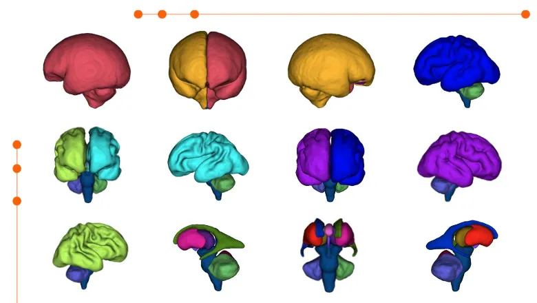
The researchers split up the different types of CHD into groups according to “cerebral substrate delivery”, i.e. how the CHD might affect the delivery of oxygen and nutrients such as glucose into the brain. They then used advanced foetal magnetic resonance imaging (MRI) techniques to show that the groups of CHD associated with lower cerebral substrate delivery tended to have smaller brains, whereas forms of CHD where cerebral substrate delivery was normal showed no differences in brain growth compared to a control group of unaffected pregnancies.
Understanding the biological mechanisms behind the impairments in brain development that we see in some cases of CHD is important for trying to decode how and why they occur, and what we might be able to do to prevent their occurrence in the first place. Information about the different types of CHD and their association with foetal brain growth helps clinicians when having discussions with women and families who are pregnant with a foetus with CHD.
Daniel Cromb, Clinical Research Fellow and PhD Student, School of Biomedical Engineering and Imaging Sciences
This research also marked a successful collaboration between researchers at King's and clinicians at Evelina London. Over 500 women consented to their MRI scans from fetal cardiology at Evelina London being used for research over a period of eight years. The process of foetal MRI scanning was overseen by clinicians at the hospital as well as researchers from the School of Biomedical Engineering & Imaging Sciences. "The process has been very helpful in highlighting the potential clinical impacts of our research", said Daniel Cromb, Clinical Research Fellow at the School.
This research highlights the amazing collaborations we have in place here at King’s College London and Guy’s and St Thomas’ Hospital, using cutting-edge prenatal imaging to understand more about how the heart and brain develop together in our patients with CHD.
Dr David Lloyd, Clinical Research Fellow, School of Biomedical Engineering & Imaging Sciences, and consultant in paediatric and fetal cardiology at Evelina London Children’s Hospital
In this story

Professor of Perinatal Imaging & Health

PhD Student