We have had a lot of help from the fantastic team at King’s Medical Engineering Quality Management System who are helping us to move the device through from a prototype to a nationally regulated device which can be used to help plan these complex procedures.
Dr Natasha Stephenson, Clinical Research Fellow in Congenital Cardiovascular Imaging at the School of Biomedical Engineering & Imaging Sciences
16 February 2022
New Virtual Reality technology to repair hearts
Immersive technology could shorten operating times and reduce the need for multiple surgeries
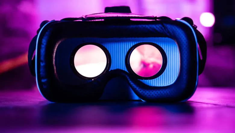
Researchers from the School of Biomedical Engineering & Imaging Sciences have developed new Virtual Reality (VR) technology, funded by the British Heart Foundation (BHF), which could improve outcomes for the thousands of patients who undergo a surgical or keyhole procedure for congenital heart disease surgery every year.
Every day in the UK around 13 babies are diagnosed with congenital heart disease - heart conditions that develop in the womb, before a baby is born. Depending on the severity of their condition, they might need one or more procedures to help their hearts function normally.
The technology, which has been developed along with researchers at Evelina London Children’s Hospital, brings together scans that are routinely used to plan congenital heart disease surgery to create a three-dimensional, beating digital double of the heart.
The researchers hope that using VR to plan and practice procedures will shorten operating times and reduce the need for multiple surgeries, leading to better outcomes and experiences for patients and their families. They hope that it could be in regular use within the next two years.
Dr Natasha Stephenson, Clinical Research Fellow in Congenital Cardiovascular Imaging at the School of Biomedical Engineering & Imaging Sciences said in a lot of way the validation process is lengthier because there is not much data about the clinical reliability of this kind of technology. Since the system us novel, proof that it works is needed and that will be safe to use to plan congenital heart procedures.
“One benefit of being co-located between King's and GSTT/Evelina is that we are embedded in the clinical team. This means it is easy to access cardiologists, surgeons and interventionalists who provide invaluable feedback and help us tailor the system to be as useful as possible for clinicians.”
Trials of an early version of the technology, which used only echocardiograms (ultrasound scans of the heart) to create the VR heart, found that surgeons preferred it for understanding the anatomy of their patient’s hearts. They also reported that it increased their confidence and improved their decision making.
Funding from the BHF has supported the researchers to add two more types of scans into the system - computed tomography (CT) and magnetic resonance imaging (MRI). While these types of scans are regularly used to help plan surgeries, they are usually only viewed on a flat screen.
This exciting project has benefitted from the input of patients and clinicians to help focus on areas that are most important to them. Initial feedback has been hugely positive. The co-location of the engineering and clinical teams allows us to rapidly respond to this feedback and develop a product that is best placed to improve patient care.
Dr Kuberan Pushparajah, Clinical Senior Lecturer in paediatric cardiology, School of Biomedical Engineering & Imaging Sciences
Surgeons using the technology are immersed into the heart. It allows them to interact with and manipulate the images however they like. They can also test options for the procedure in VR before they get to the operating table.
Every patient with congenital heart disease has their own set of unique changes to their heart. By giving surgeons a better understanding of this and offering them an opportunity to practice and perfect operations, the researchers hope this technology will also help to improve the experiences of thousands of patients and their families each year.
Procedures to repair the heart’s anatomy can be complex, and surgeons don’t like surprises. Our technology will allow surgeons to plan and practice these procedures, and we’re currently applying for approval for it to be used in this way. “We think that this technology could also be used outside of congenital heart disease surgeries, to plan any procedure which aims to correct a structural problem within the heart, such as valve surgery in an adult patient.
Lead researcher Professor John Simpson, Professor of Paediatric and Fetal Cardiology at Evelina London and King’s College London
Congenital heart disease is the most common cause of birth defects in babies born in the UK. Every year thousands of heart operations and other procedures are performed for children and adults with congenital heart disease to stop them developing heart failure. Some people will need several procedures during their lifetime. This new technology could help to make congenital heart disease surgery even more successful. It could also support people to better understand the heart or blood vessel abnormalities they are born with and what is being proposed to mend them, which can be empowering for people living with congenital heart disease.
Dr Sonya Babu-Narayan, Associate Medical Director at the British Heart Foundation and consultant cardiologist
The technology has also received significant funding from Evelina London Children’s Charity.