06 June 2019
Nikon Imaging Centre at King's established as the Nikon super resolution microscopy centre for Europe
Dr Katherine Lau, Product Manager for Super Resolution Microscopes, Nikon Instruments Europe B.V.
Nikon Instruments Europe is excited to further their existing relationship with KCL by establishing the Nikon Imaging Centre (NIC) at King’s as a Nikon super resolution microscopy centre for Europe
Bio-imaging is a powerful tool in biological research, providing valuable spatial, quantitative and dynamic information on biological molecules. This information enables researchers to investigate the structure, functions and mechanics of a wide range of biological molecules.
The NIC at King’s College London was established in 2012 with the aim of providing the most cutting-edge imaging technologies to researchers in the biological sciences. By establishing the NIC at KCL, Nikon are able to engage with researchers, understand their imaging needs and stay in touch with the latest imaging challenges they face. This relationship enables Nikon to provide innovative technologies which meet the academic community’s needs.
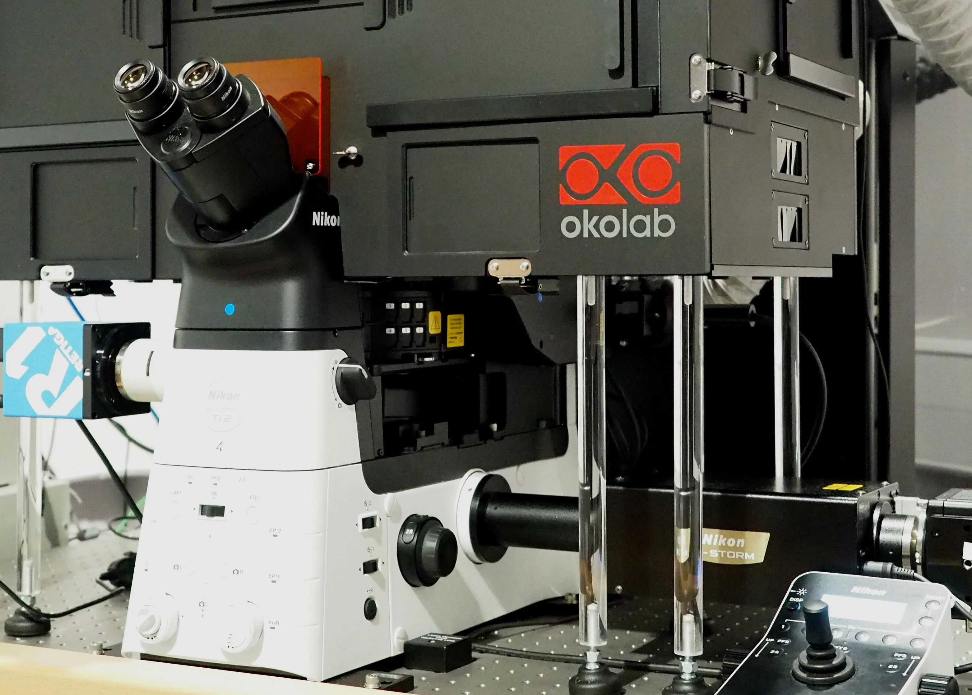
Nikon Instruments Europe is excited to further their existing relationship with KCL by establishing the NIC at King’s College as a Nikon super resolution microscopy centre for Europe, enabling KCL researchers to go beyond the confocal spatial resolution limit. The latest generation of super-resolution microscopes, the N-SIM S and N-STORM 5, were installed at KCL in late 2018 and early 2019, respectively. N-SIM S provides XY spatial resolution of 115 nm in 2D- and 3D- SIM modes, and 86 nm in TIRF-SIM mode. The system is ideal for live cell imaging, allowing more structural dynamic subcellular details to be captured. With N-STORM 5 researchers can achieve approximately 20 nm XY resolution and 50 nm Y resolution. Such superb resolution enables researchers to study their biomolecules of interest on a single molecule level.
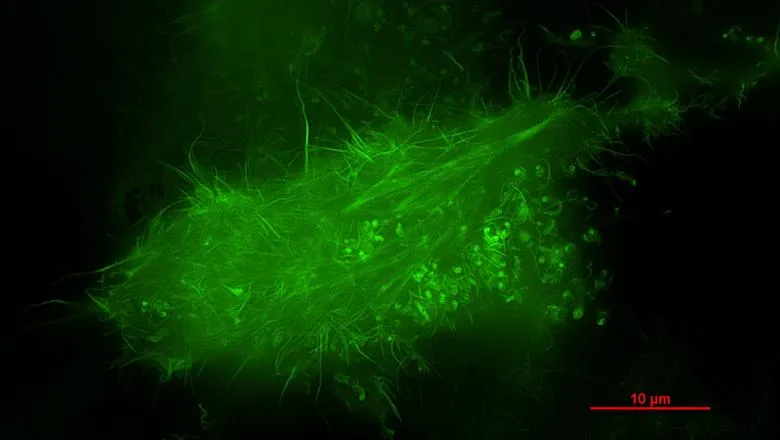
Tommy Pallet, a PhD student at the Randall Centre for Cell and Molecular Biophysics, KCL commented: “Imaging tiny and highly dynamic structures such as the cell skeleton has always been a challenge for biologists. My research focuses on developing new microscopic techniques to study the structure and function of a specific part of the cellular skeleton: the actin network. This network is of particular interest since it is the driving force behind cell movement, which is central to many biological processes such as neural development, wound healing, and cancer metastasis. Actin makes up the finest of the networks, and so high resolution is paramount, and the network is regulated by a whole host of different molecules, so multiple labelling (and multi-channel imaging) is also extremely desirable.
Excellent resolution can be obtained with electron microscopes, but it is difficult to preserve structures of interest during preparation, difficult to investigate different structures or molecules simultaneously and analyse them, and impossible to extend experiments into a live cell environment. The new N-STORM 5 system is critical to circumventing these issues. Using super-resolution optical microscopy, it is possible to probe different structures and molecules simultaneously whilst still achieving a resolution close to that of an electron microscope. We are also investigating the potential of taking this into living cells, which would enable us to study the network in action, which is extremely important when researching cell migration – an intrinsically dynamic process.”
Daniel Matthews, the NIC Facility Manager commented: “The partnership with Nikon means that that we can offer the latest optical microscopy technology to users of the centre. In addition to providing instrumentation Nikon closely supports activities at the NIC, helping to run hands-on workshops and offering advice on microscope setup and data processing.”
For further information about the NIC please see: https://nic.kcl.ac.uk/