05 December 2023
Odontodes: The Developmental and Evolutionary Building Blocks of Dentitions
Professor Moya Meredith Smith and Dr Aaron LeBlanc contribute to a newly published book on the odontode system.
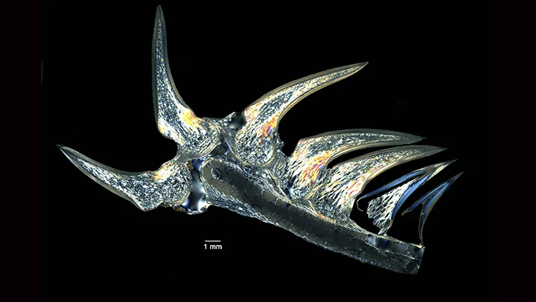
The odontode system, which encompasses teeth and other dentine-based structures, is ancient. Odontodes are present in the oldest vertebrate fossils, dating back 500 million years, and still play an important role in the anatomy and function of living jawed vertebrates. Fossils preserve odontode tissues with remarkable nanoscale fidelity, allowing the evolution and diversification of the odontode system to be studied in deep time as well as across the diversity of living vertebrates. This synthetic volume presents an overview of odontode research by internationally leading researchers from different fields of biology.
Professor Moya Meredith Smith and Dr Aaron LeBlanc from the Faculty of Dentistry, Oral & Craniofacial Sciences have contributed chapters to this book.
Moya Meredith Smith’:
Chapter 5: Odontoblast Repertoire Delivers Significantly Different Dental Tissues from Pluripotent Neural Crest-Derived Cells
Authors: Moya Meredith Smith, Aaron R. H. LeBlanc, Charlie Underwood, and Zerina Johanson
This chapter, led by Moya Meredith Smith from FoDOCS, is a chapter in the Taylor and Francis book ‘Odontodes’ in a prestigious series on evolution to be published on December 8th. This chapter presents dentine as the quintessential cell product in odontodes (teeth and tooth-like organs) and a universal tissue found in all tooth types. A common feature is that the dentine-producing cells (odontoblasts) are retained for the life of a tooth and remain in the soft tissue of the pulp, allowing them to repair damaged dentine. Our focus is on their unique morphology, where each odontoblast has a single cytoplasmic process embedded within a tubule of the mineralised tissue and reaching from the junction of the first formed organic matrix to the pulpal surface. This feature tracks the journey cells take over their entire life of tissue production, being easily recognizable in developing and adult tissues, extant and fossil.
Odontoblasts are not only critical for maintaining a tooth, but for vital tissue repair through their migratory and invasive properties. This chapter demonstrates their ancient reparative abilities within the odontode armour of fossils of jawless vertebrates. Moreover, odontoblasts are present not only in teeth but also in dental plates of some of the most unusual, toothless fishes: chimaeras. This pluripotent repertoire makes odontoblasts, as cells derived from cranial neural-crest (CNC), unique among all mineralised tissues. The migration of odontoblasts, recorded in the hard tissues they leave behind, is fundamental to the role and function of this universal tissue as a responsive and sensitive component of any tooth. We have been able to show the migratory pathways of these odontoblasts in all varieties of dentine in fish both living species and the related fossils using richly illustrated plates and figures. Using the distribution of the tubules, as indicators of living cell history we have been able to demonstrate the variations in different species, ranked in phylogenetic order, giving a living picture of the cellular function of odontoblasts with a varied repertoire derived from their migratory pathways, imbedded in their embryonic inheritance.
Dr Aaron LeBlanc:
Chapter 6: Shifting Perspectives in the Study of Amniote Tooth Attachment and the Path Forward to Establishing Vertebrate Periodontal Tissue Homology
Author: Aaron R. H. LeBlanc
This chapter by Aaron LeBlanc (FoDOCS) tackles a 150-year-old problem: what do we call the supporting tissues of the teeth in non-mammalian species? While we have concrete developmental, histological, and anatomical definitions for the human periodontal tissues – root cementum, the periodontal ligament (PDL), and alveolar bone – this vocabulary breaks down as researchers work on progressively more distantly related vertebrate species. This problem goes beyond the terminology and affects our ability to study the evolutionary origins of our periodontium from our more “reptile-like” ancestors. The chapter addresses this by refining our definitions of the periodontal tissues through a comparative histological approach. Using detailed colour images, LeBlanc illustrates what the cementum, PDL, and alveolar bone of mammals, crocodiles, lizards, snakes, dinosaurs, and other extinct animals look like under the microscope, providing a framework for identifying them in thin sections of modern and fossil animals alike. The chapter uses case studies from previous research by LeBlanc and others to show that this comparative approach reveals a surprisingly complex evolutionary history of the periodontal tissues. While cementum, the PDL, and alveolar bone are ancient features of teeth, the capacity for the periodontium to either completely mineralize or retain a soft, ligament-supported tooth attachment has changed many times throughout the evolutionary history of different groups of reptiles and synapsids (the larger group to which mammals also belong). This chapter ends by laying the groundwork for future studies of the periodontal tissues in fish, sharks, and the most ancient tooth vertebrates.
In this story

Professor of Evolutionary Dentoskeletal Biology
Lecturer in Dental Biosciences