“It was indeed a great honour to have been recognised by the Royal Photographic Society and the Royal Colleges with this award for our work. Advanced imaging technologies are critical in our healthcare today. From artificial intelligence enabled technologies to precise, data-rich medical scans and digital models, the impact of these developments are far reaching for clinicians and patients.”
Professor Reza Razavi
14 November 2019
Professor Reza Razavi honoured for work in medical imaging
Professor Razavi was awarded the Combined Royal Colleges Medal
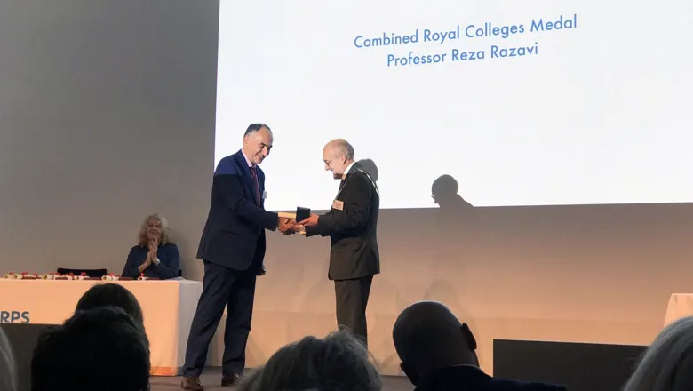
Professor Reza Razavi was last night awarded the Combined Royal Colleges Medal with Honorary Fellowship for outstanding contribution to medical imaging and its application in the service of medicine and surgery at the 141st Royal Photographic Society’s Awards.
The prestigious awards were established in 1878 to celebrate outstanding contributions to photography and now span the fields of art, science, education, film and publishing. The combined Royal College’s Medal was established in 1961 is awarded jointly with a number of the Medical Royal Colleges including The Royal Colleges of Physicians and Surgeons.
Holding senior leadership research positions at King’s College London, King’s Health Partners and as consultant cardiologist at Guy’s and St Thomas’ NHS Foundation Trust, Professor Razavi said the awards showcase innovative research in medical imaging and enable its importance to be communicated to the public.
Professor Razavi’s prize follows a number of successes he and his team have achieved during the last 12 months, including a grant award from Innovate UK to use artificial intelligence tools and imaging data for value-based healthcare.
Earlier this year, Professor Razavi and his team spearheaded revolutionary new technology for detecting heart defects in babies. The technology produces a 3D computer model of unborn babies' hearts from different angles and a MRI scan takes hundreds of photographs inside the womb.
Thanks to this new approach, specialists are now able to diagnose congenital heart disease in unborn babies earlier and safer.
Professor Razavi’s group was also the first to perform MRI guided cardiac catheterisation in patients and more recently carried out entirely MR guided cardiovascular interventions.
Key areas of focus for the team are cardiac imaging in relation to congenital heart disease, electrophysiology and heart failure, image guided intervention, XMR (X-ray and MRI) guided cardiac catheterisation and methodological advances to move to faster 3-Dimensional cardiac imaging.