14 July 2021
Real-time surgical hyperspectral imaging system for safe tissue removal undergoes clinical feasibility study
Researchers from the School of Biomedical Engineering & Imaging Sciences at King’s College London, in collaboration with researchers at the Research in Orthopedic Hospital (ROCS), University Hospital Balgrist, University of Zurich, Switzerland, have conducted a feasibility case study of a novel surgical hyperspectral imaging research system. The outcome of this research study further bolsters the potential and directly informs the ongoing development of this new technology which could lead to increased surgical precision and patient safety.
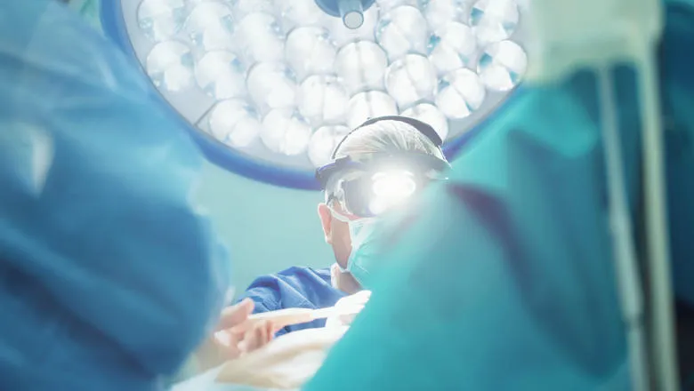
This clinical feasibility case study was published in a special issue of Journal of Physics D: Applied Physics. Its results demonstrate that the research system seamlessly integrates into an operating workflow, with minimal disruption to surgery. The clinical feasibility case study was conducted with a patient undergoing a spinal surgery. An ex vivo study was also performed. Each study provided crucial insights to develop a system suitable for acquiring high-quality data in a highly dynamic and constrained environment as it is in an operating room during surgery.
These findings pave the way towards further system development and clinical validation to assess its potential clinical benefit for patients.
Researchers say the key outcome of safe use and seamless integration into a surgical workflow indicates the system’s viability for use during surgery – the very purpose for which it was designed.
Dr Michael Ebner, one of the lead researchers of the study, said with these findings and the ongoing lab development of the prototype system, they can now advance developing real-time surgical guidance.
"This initial pilot study was a key step to demonstrate a safe surgical system can be developed to provide critical, but currently unavailable, information to the surgeon for improved patient outcomes.”
Dr Jonathan Shapey, Clinical Senior Lecturer in Neurosurgery, who led the preclinical development of the system and operated the device during the surgery together with colleagues at University Hospital Balgrist, University of Zurich, said:
“Existing systems do not allow for seamless integration into the surgical workflow nor do they provide real-time instantaneous feedback crucial to support effective decision-making during surgery. In contrast, our real-time hyperspectral imaging uses a smart camera technology with the potential to transform the way surgeons can visualise tissue during surgery. By highlighting different tissue types, for example by differentiating tumour from healthy tissue live and in real-time, HSI could significantly improve the safety and success of surgery.”
The goal of the researchers is to develop a system that provides clinicians with augmented vision for clear visualisation of vital tissue information throughout surgery. This technology could provide critical information to increase both surgical precision and patient safety for improved surgery and patient outcomes.
Professor Tom Vercauteren, PI of the study, emphasised the importance of multi-centre research and industrial partnerships for successful development of new surgical technology:
“This study has been pivotal in many ways. The success of this new cross-institutional collaboration served as a steppingstone to secure joint follow-up funding as part of the EU-funded H2020 Functionally Accurate Robotic Surgery (FAROS) project. It also gave us confidence about the industrial potential of the technology.”.”
Professor Philipp Fürnstahl, University of Zurich highlighted the breadth of potential applications of this technology:
“Hyperspectral imaging opens up new research possibilities in the field of computer-aided surgery. It may become an exciting intraoperative technology to enhance the surgeon’s view on crucial anatomical structures.”
Besides the provision of precise margin information of tumour and its relation to healthy tissue structures, a surgical hyperspectral imaging system could for example also provide blood flow and perfusion understanding.
Leveraging the result of this study, Hypervision Surgical was spun-out from King’s College London by Michael Ebner, Tom Vercauteren, Jonathan Shapey and Sebastien Ourselin. The company will further develop this promising technology into a commercial medical device for the patient benefit. Dr Ebner was also the recipient of an Enterprise Fellowship from the Royal Academy of Engineering to support the development of this novel lightweight camera system.
In this story

Professor of Interventional Image Computing

Research Associate