To be able to have tools that help increase the diagnostic confidence so that women can be counselled more accurately is very important
Jackie Matthew, NIHR Clinical Doctoral Research Fellowship recipient
16 July 2020
Research Sonographer, Jackie Matthew awarded prestigious NIHR fellowship award
Researcher at School of Biomedical Engineering & Imaging Sciences awarded NIHR Clinical Doctoral Research Fellowship award
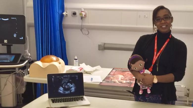
Jackie Matthew at the Centre for the Developing Brain at The School of Biomedical Engineering & Imaging Sciences has been awarded the prestigious National Institute for Health Research (NIHR) Clinical Doctoral Research Fellowship award.
A Research Sonographer and Radiographer, Jackie Matthew will complete her PhD and clinical academic training in new antenatal 3D ultrasound and MRI technologies to more accurately assess facial and cranial features for potential congenital anomalies.
Clinical Challenge
New methods of accessing and reviewing imaging data could better equip clinicians to offer to parents more accurate counselling and follow up care.

Despite 3D ultrasound scans being popular amongst parents in private ‘baby bonding’ clinics, the technique is not used often clinically for diagnosis as it is time consuming to perform, requires specialist training and the images are often partially obscured because of the baby’s position or movements.
“In some facial and cranial anomalies in particular, the antenatal detection rate remains quite low,” Ms Matthew said.
“Therefore, improvements in the 3D technology would be beneficial as it can negate some of the difficulties that occur when looking at the curved structures of the face and head in two dimensions.”
For example, cleft palate, resulting in a gap at the roof of the mouth, and craniosynostosis, early fusion of the cranial sutures, can have a significant impact on the baby’s health and often requires surgical treatment, however it can difficult to diagnose when baby arrives resulting in a delay to treatment.

In these situations, antenatal diagnosis would help.
The Technology
“Using 3D reconstructions from ultrasound and MRI could also allow clinicians to assess minor changes in facial appearance that are not necessarily overt structural anomalies but features that could be part of a genetic syndrome,” Ms Matthew said.
She said many rare conditions include facial features that usually go unrecognised on antenatal imaging but if identified would aid the diagnostic work up, for example, the identification of low set or poorly formed ears is seen with many syndromic and genetic conditions such as Down’s syndrome, Turner syndrome and Beckwith-Wiedemann syndrome.
"It is important that the research looks at the role of MRI as well as ultrasound, because less is known in terms of how well MRI can assess minor craniofacial anomalies using 3D reconstructions."
With the development of MRI slice to volume reconstructions by King’s researchers, 2D MRI scans can now be converted to motion corrected 3D volumes.
This means that clinicians can still look at the images in 2D slices, but now they can look at the anatomy from any 2D angle and they have the potential to build 3D surface models, meaning previously inaccessible anatomy could now be seen well.
Parents
“If you’ve been given the all clear during your routine screening and there’s no suspected anomalies on the ultrasound scan, and then baby arrives and there are anomalies - that can be distressing for parents.”
“From the parents’ point of view, they may wonder why it wasn’t seen sooner especially if any delays in diagnosis may have affected baby’s health. Being able to pick things up earlier means that those delays in the care management or treatment, which could impact mum’s or baby’s health, could be avoided."
Jackie’s work has roots in the intelligent Fetal Imaging and Diagnosis (iFind) project, which investigates the use of engineering developments including artificial intelligence and machine learning, robotics and multiple ultrasound transducer technology, to improve the way fetal anomalies are detected using novel tools that aren’t yet used routinely in NHS scan clinics.
Ms Matthew concluded, “Ensuring the voices of parents are included in the development of new scanning tools is very important to me so that their needs and concerns can be addressed in this and in future related research. I have a number of focus groups planned with parents and I have just launched social media pages to allow me to share details of this project and connect with parents.”
You can keep up to date with Jackie’s project by following her on Twitter, Instagram, or by liking her Facebook page.