This successful collaboration between experts in biomaterial science and stem-cell technologies really demonstrates the impact and importance of interdisciplinary collaboration. I’m very excited to be working at the interface of physical and life sciences and look forward to the future developments and creative new applications of this work.
Aimee Cheesbrough, PhD student at UCL
21 March 2022
Researchers create synthetic scaffold to help grow lab muscle tissue
The nanofiber scaffold helps the growth of synthetic muscle tissues from stem cells, allowing scientists to understand the mechanisms of muscle diseases and test new drug candidates.
The Lieberam Lab, in collaboration with the Song Group at UCL and led by PhD student Aimee Cheesbrough, have developed a new synthetic scaffold to grow muscle cells from human stem cells, allowing scientists to effectively grow synthetic muscle tissue for research into muscle diseases.
The results, published in Advanced Materials, showed that the scaffold successfully grew skeletal muscle tissue.
Muscles are attached to their corresponding bone by tendons, and muscles contract to move a particular part of the body.
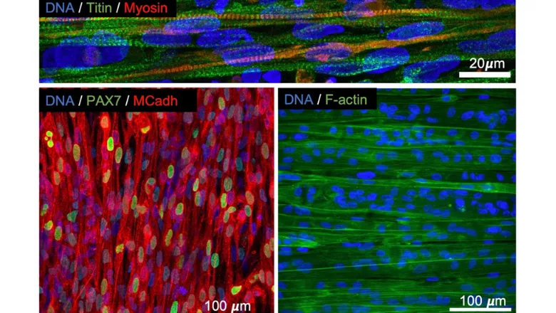
Current research into muscle diseases has a problem with growing muscle tissues for experiments. This is because the myofibers (muscle cells) that collectively form the muscle tissue are surrounded by the extracellular matrix (ECM) which is difficult to replicate in laboratory conditions.
The ECM acts as a connective scaffold that helps form the structure of myofibers. Having the ECM scaffold in the environment is vital for the growth and maturation of muscle tissue. Without a reliable way of replicating the ECM, scientists are unable to grow muscle tissues.
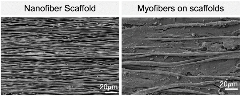
The new research explores the development of a synthetic, flexible scaffold which replicates the ECM. Electrospinning technology weaves elastic nanofibers from synthetic biomaterials to build an ECM-replicant scaffold that can be used to grow muscle tissue from human stem cells.
The paper shows that the synthetic-ECM – the nanofiber scaffold – successfully supported the normal growth and stabilisation of skeletal muscle tissue.
The authors also inserted a molecular light sensor into the genome of the stem cells to create myofibers that contracted upon exposure to light pulses. This means artificial muscle tissue can be contracted like muscles in the body.
As a result, scientists can create many identical muscle tissue models for experiments. These help researchers to better understand contraction in muscle diseases, as well as testing drug candidates with the same ideal muscle tissue model.
As well as modelling and testing drug candidates for muscle diseases, the artificial ECM scaffold has the potential to be used to grow replacement muscle tissue for patients with skeletal muscle injuries.
The researchers also noted that the synthetic muscle tissue can be used to study a range of diseases. Peter Harley, a PhD student of the Lieberam Lab, has been growing myofibers that are connected to motor neurons made from human stem cells. This is designed to mimic a neuromuscular circuit for the study of Amyotrophic Lateral Sclerosis (ALS).
In this story

Senior Lecturer