This work shows promise in identifying a technique for continuous monitoring of perfusion and oxygenation of a transplanted kidney. This method may be applicable to other organ types and also illustrates the importance of interdisciplinary collaboration to address important health issues.
Mr Pankaj Chandak, transplant research fellow from the School of Immunology & Microbial Sciences at King's
28 April 2025
Researchers develop model for real-time monitoring of kidney transplants
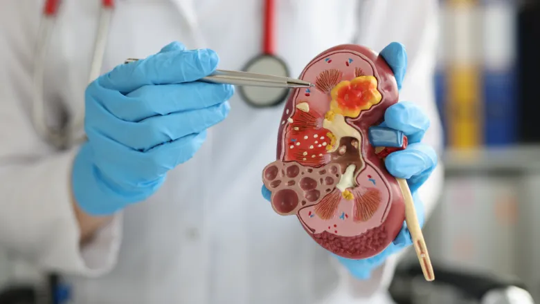
Researchers at King’s College London, the University of Bristol and Great Ormond Street Hospital have developed a model using human kidneys and warm machine perfusion that could allow for real-time monitoring of kidney transplants.
Co-authored by Mr Pankaj Chandak, a transplant research fellow from the King’s School of Immunology & Microbial Sciences and Dr Danothy Bennett from the Interface Analysis Centre at the University of Bristol, the study published by The Royal Society aims to enhance post-operative care by developing a system that could potentially monitor perfusion of transplant organs in real time, allowing for earlier intervention if complications were to arise.
A transplant is one of the main treatments for end-stage organ disease, but there is a risk of post-operative complications, such as poor transplant blood flow, that may result in loss of the organ if timely intervention is delayed.
Currently, kidney transplants are monitored by clinical assessment of the patient with blood tests and, if needed, relevant imaging such as ultrasound scans. However, these methods have limitations, including delayed detection of complications and reliance on direct access to imaging facilities, which may be an out-of-hours service and not always readily available.
A research team, supervised by Professor Nizam Mamode from King’s, Dr Wesley Hayes from Great Ormond Street Hospital, and Dr John C.C. Day from the Interface Analysis Centre at the University of Bristol, has developed a proof-of-concept technique to continuously monitor kidney oxygen levels in real time.
The team used human kidneys from NHS Blood and Transplant that were not suitable for transplantation and that were offered for research purposes. These organs were then placed on a specialised bypass machine that simulates the physiological conditions required for an organ to function outside the human body and maintain metabolic activity (known as ex vivo normothermic machine perfusion).
Special custom-made probes that use light to measure properties of a sample (spectroscopic optical reflectance probes) developed by the team at the Interface Analysis Centre at the University of Bristol were then placed on the surface of the kidneys to measure oxygen levels in real time whilst the organs were undergoing warm machine perfusion.
To validate the technology’s effectiveness, the researchers, using the bypass machine, simulated real-world transplant complications such as thrombosis (blood clot), rejection, and blood loss. The probes successfully detected oxygenation fluctuations in response to these conditions, demonstrating their potential for clinical application.
This method of using special optical sensors to detect dynamic changes in oxygenation in the organs could help medical teams to quickly detect and address blood flow problems, offering an additional method of monitoring to currently available methods. With further work, the researchers foresee potentially integrating reflectance probes into post-operative care by placing them alongside surgical drains, allowing continuous monitoring of graft perfusion in the days following transplantation.
This work highlights the feasibility of real-time, continuous monitoring for kidney transplants and paves the way for further research into broader clinical applications.
The research was supported by funding from multiple sources. See full details here.
For more details, you can read the full study published in Royal Society Open Science.
In this story

Research Fellow