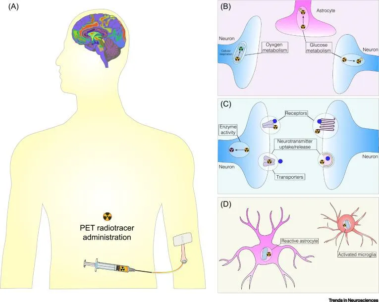
PET could answer these questions more specifically. We can address specific questions like whether certain neurotransmission pathways are being affected or other specific types of cells such as astrocytes and microglia.
Mr Igor Fontana, PhD Student
13 November 2020
Researchers investigate possible link with COVID-19 and neurological damage
There is a growing body of literature looking into the potential link and how PET imaging could be used
Recent studies have shown that a substantial number of COVID-19 patients develop neurological manifestations. Nevertheless, pathological changes in the central nervous system induced by SARS-CoV-2 are still uncertain.
In a new paper published in Trends in Neurosciences, Positron Emission Tomography (PET) researchers from the School of Biomedical Engineering & Imaging Sciences in collaboration with the Universidade Federal do Rio Grande do Sul (UFRGS), Brazil, suggest that PET imaging could be useful to address potential brain changes in COVID-19 patients.
The researchers suggest that PET radiotracers, molecules that have high specificity towards a biological target, may reveal “where” (brain regions and cell types affected) and “how” (changes in neurotransmission, physiological and metabolic parameters) SARS-CoV-2 affects the brain.
In addition, early PET scanning could help indicating “when” a patient is at risk of developing neurological manifestations due to COVID-19.
Published in Trends in Neuroscience, the paper also discusses very recent findings demonstrating the applicability of widely applied PET radiotracers in clinical research.
The PET radiotracer [18F]FDG, thought to reflect synaptic dysfunction, loss of connection between cells, indicated regions affected in the brain of COVID-19 patients.
Lead researcher PhD student Igor Fontana says that there is still much to learn about the precise location of these manifestations and how specifically they affect the brain.
"Other neuroimaging techniques like MRI are being applied on larger cohorts, but there are still a lot of studies that need to be done,” he said.
"There are just a few papers talking about PET imaging in the brain of COVID-19 patients and the notion that neurological manifestations are linked to COVID-19 is even more recent.
In recent correspondence to the Lancet Neurology, clinicians in Jerusalem reported the case of a 45-year-old man with confirmed COVID-19 infection who developed secondary Parkinsonism due to SARS-CoV-2.
A precise diagnosis could be only made with the application of brain PET imaging.
“This is all new and the neurological manifestations are becoming more eminent – there are new pathways and ells that can be affected. If neurological changes evolve into a chronic state, we should look towards the development of neurodegenerative diseases,” Mr Fontana said.
The researchers said while future research could investigate pharmacological interventions, immediate focus is to be on how COVID-19 specifically affects the brain.