Now more than ever, we need to support our early career researchers. This scheme aims to ensure a clear path for outstanding candidates towards obtaining external fellowship funding which would facilitate a long-term future at King’s under our new King’s Fellows scheme.
Professor Reza Razavi, CME Director
28 January 2021
Researchers receive CME Research Fellowships for outstanding postdoc scientists
The CME scheme supports postdoc researchers to obtain external funding

Researchers from the School of Biomedical Engineering & Imaging Sciences part of the Wellcome/EPSRC Centre for Medical Engineering (CME) have been awarded CME Research Fellowships that support outstanding post-doctoral scientists for a period of independent working to obtain external fellowship funding.
The Fellows were selected on the scientific merit of the research project, feasibility of their proposal and the potential to successfully apply for external fellowship funding within the first 6 months of their CME fellowship.
Congratulations to the CME Fellowship Awardees. One of the key aims of the Centre is to support our Early Career Researchers and with the Centre’s support we hope to ensure ongoing success for these talented researchers.
CME Centre Manager, Dr Shalini Jadeja
“The three recipients exhibited tremendous long-term potential with their research and we are excited to watch the research grow and researchers succeed at King’s,” he said.
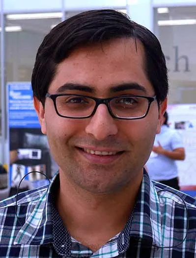
One recipient Dr S.M.Hadi Sadati is looking at reinventing catheter steering with soft robotics: a tele-supervised semi-Autonomous wound free Robotic Thrombectomy in acute stroke (ART).
The most common underlying causes of stroke are cardiac rhythm abnormalities and carotid artery disease where blood clots are generated which can block brain arteries.

Recent evidence shows that if mechanical devices are used to unblock the brain artery within 6 hours of onset of a blockage, more patients are independent after treatment, not requiring support for everyday living.
But few operators have the expertise to perform the unblocking procedure (78 in UK) and these experts are concentrated in a small number of specialist centres (24 in UK) to allow a continuous daytime service.
Dr Sadati’s ART project aims to develop a robotic unblocking procedure to allow local treatment to be performed remotely from a specialist neuroscience centre to reduce the time gap from diagnosis to the procedure having huge impact in patient benefit.
“ART explores novel ideas following the identified shortcomings in catheter interventions for inclusion, navigation, intervention, and user-study,” Dr Sadati said.
ART can be useful for other aligned applications such as cardiac ablation, renal stone removal, and shunt introduction. The proposed incision method can be beneficial in safe navigation of vital organs in brain and spine minimally invasive surgery.
Dr S.M.Hadi Sadati

Dr Laura Peralta Pereira’s project proposes a new ultrasound approach: Hyper-aperture ultrasound (HAP-US) where multiple, flexibly positioned, small probes are combined to operate as a larger, flexible and multi-view aperture.
While ultrasound can provide real-time information through a portable, low-cost, painless and radiation-free method, there are still some key challenges that limit the usability of conventional ultrasound.
Dr Peralta said medical ultrasound images are blurred (low resolution) and have a restricted field of view that often prevents visualization of the entire organ of interest.
“Creating such a large aperture could lead to significant improvements in fundamental ultrasound features, enhancing not only diagnostic imaging, but also benefiting all methods that reply on ultrasound imaging with significant clinical applications such as blood flow measurements or image-guided tasks,” Dr Peralta said.
My aim is to develop the core methods required to establish HAP-US as a fully operational technology and provide evidence of its powerful capabilities. Successful development and demonstration of HAP-US throughout this research could eventually greatly expand the clinical opportunities of the ultrasound.
Dr Laura Peralta Pereira

Dr Yijing Xie, L’Oréal UK&I and UNESCO for Women in Science Rising Talent Fellow is developing a functional optical imaging system for surgical guidance.
Dr Xie will engineer two emerging imaging modalities, light field imaging and multispectral imaging, into one compact device and develop a complex imaging reconstruction algorithm.
“It will be a world first cost-effective 3D multimodal exoscope designated for use in the surgical environment,” Dr Xie said.
Dr Xie said this imaging system is able to provide brain functional information and render it in to a reconstructed 3D surgical field enabled by the featured light field technology.
Primarily used in the awake craniotomy for brain tumour resection, a surgical technique used to remove brain tumour that is adjacent to brain regions with critical motor, language or cognitive functions, it will inform surgeons a classified and labelled brain tissue map, to differentiate tumour tissue for precise removal.
The surrounding functional brain will be identified and preserved, minimising the risk of causing post-surgical neurological deficit, such as the inability to speak, if a brain region of language function is damaged.
“This way the tumour tissue can be maximally removed and important brain functions will be protected,” Dr Xie said.
Patients will have prolonged survival benefiting from a maximal tumour removal, and better quality of life because of reduced post-surgical deficits.
Dr Yijing Xie
