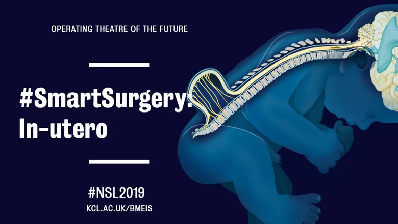
The Operating Theatre of The Future at New Scientist Live
Come visit The School of Biomedical Engineering & Imaging Sciences at this year's New Scientist Live, October 10-13
03 October 2019
A team of researchers at King’s College London School of Biomedical Engineering & Imaging Sciences are proving the impossible with their breakthrough technologies for fetal therapy and surgery.
A team of researchers at King’s College London School of Biomedical Engineering & Imaging Sciences are proving the impossible with their breakthrough technologies for fetal therapy and surgery.
Their work takes place in a large research programme called Guided Instrumentation for Fetal Therapy and Surgery (GIFT-Surg) which is developing the technology, tools and training necessary to make fetal surgery a viable possibility in the treatment of congenital birth defects, such as Spina Bifida, Twin-to-Twin Transfusion syndrome, Congenital Diaphragmatic Hernia and lower urinary tract obstruction.
In collaboration with KU Leuven in Belgium, University College London, Great Ormond Street Hospital, University College London Hospital NHS Foundation Trust and UZ Leuven, GIFT-Surg comes from the simple analysis that nowadays every pregnancy is being followed very thoroughly and we have very accurate ways of detecting any anomaly or any disease in fetuses long before birth.
But once this is done and if clinicians have a diagnosis, then the therapeutic steps are often very limited. There’s a discrepancy between what pathologies can be identified and how plausible it is to act.
“Looking at what was already out there, one of the areas that was very interesting from an engineering perspective as well, was the space of fetal surgery which has strong potential for patients but is also very challenging for clinicians,” Professor Vercauteren, co-investigator of GIFT-Surg said.
“We thought if we can design tools that can help facilitate surgeries and make them easier, then we’d be providing more options to parents in need.”
One of the clinical questions the team is trying to support with the tools they’re developing is to help parents make more informed decisions about the future of their babies.
These parents are in the stressful situation of carrying a child with abnormalities, often asking do they wait and have the disease treated after birth – which would be the natural course of operation – do they terminate the pregnancy if the severity of the disease is really complicated, or do they go for in utero therapy in some of the eligible cases?
The choice is of course difficult, especially if the parents don’t really know what the impact of the condition is for the baby.
The team has been developing ways to better look at the brain of the babies before and after surgery to potentially get better information of what the neurological outcomes of the surgery would be.
This information is relayed back not only to the surgeon and clinical team but also to the parents to help them in their decision-making process.
With these types of surgical investigations, there might be an invasive procedure done whereby a needle is inserted into the womb to take fluid or placenta tissue out to then analyse.
It’s a challenging procedure where the clinician would hold an ultrasound probe which is on the belly of the mother and with the other hand, hold the needle. The two must be aligned so the clinician can see the needle in the ultrasound image.
If it sounds complicated, it’s because it is. If things are slightly misaligned, it’s easy to confuse the tip of the needle with its shaft. For one tool, integrating several technological blocks developed by colleagues within GIFT-Surg, the team is supporting the development of a kind of GPS for ultrasound-based needle procedures where a sensor is inserted within the needle itself and where there is an active communication between the needle and the external ultrasound probe. This way, the position of the needle can be located and displayed within the ultrasound image which is provided to the clinician.
It allows the clinician to see the needle when it’s there but also not get confused between the shaft and the tip and therefore really be much more accurate in the targeting of the needle procedures.
At another end of the healthcare spectrum, Professor Vercauteren is also designing tools and image computing solutions to support better resection of tumours.
For instance, one of the key challenges in many of the tumour related neurosurgery procedures is to identify the boundaries of tumours.
It’s always a trade-off between how much can be removed to get as much tumour out as possible but being sure that there is as little impact as possible on the neurological outcomes for the patient. Seeing that boundary during surgery with the naked eye is very difficult. Machine learning tools are needed to extract, in real-time, the relevant information from various imaging modalities and present this in an actionable manner to the surgeons.
It’s something that can make a lot of difference and it can only be done with modern machine learning tools.
Professor Vercauteren wants to drive change towards a more integrated procedure where machine learning and AI agents help to support clinical staff and patients to have a much better understanding of what surgery is about, and how to deliver the right tool at the right moment to the surgeons.
"I think this is where we can really make an impact if we manage to design the algorithm that can act as a surgical assistant and really support the clinical team as needed at any given moment.”
While every day, researchers develop new methodological approaches and new technology, very often these prototypes can’t be used on patients. What’s left are exciting and interesting ideas that can only be useful if someone else takes them forward and implements them as clinical tools.
The classical researcher's perspective, explains Professor Vercauteren, is that this is the role of industry: if there’s an interesting idea that has commercial viability then of course they are going to take that technology and develop a product out that everyone will benefit from.
But it often doesn’t work quite like this. Industry usually needs proof the technology could work in patients.
“That’s why we have this gap between where the research usually ends and where the industry is usually picking it up to translate into commercial products,” Professor Vercauteren said adding that this inevitably adds a bit of waste. He is interested in exploring how this gap could be bridged.
“Of course, we’re not going to replace the industrial partners – that's not at all the case – we need commercial partners because they provide the only viable route for clinical adoption. But maybe it’s our responsibility to push the more exciting technology one step further and demonstrate their usefulness in patients.”
When it comes to GIFT-Surg, for instance, the clinical drive from surgical teams has been overwhelmingly positive, with Professor Vercauteren often being asked to expedite the development process.
“By thinking about clinical adoption from the onset of our projects and designing dedicated processes to support clinical translation of our work, I am confident we can accelerate and increase the conversion of interesting engineering ideas into clinical tool with positive impact for patients” said Prof Vercauteren.
Professor of Interventional Image Computing
Come visit The School of Biomedical Engineering & Imaging Sciences at this year's New Scientist Live, October 10-13