
New Scientist Live 2019
Be part of the world's greatest science festival
09 September 2019
Dr Rachel Sparks is working on EpiNav, pioneering software helping neurosurgeons plan keyhole neurosurgery quicker and safer.
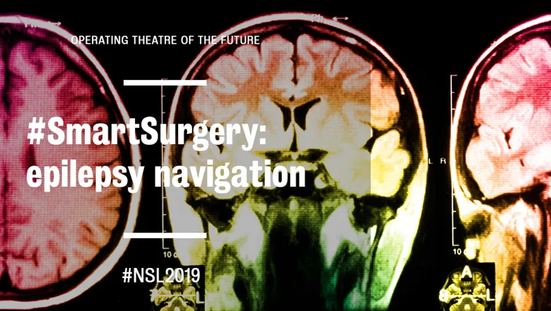
When it comes to planning keyhole neurosurgery for epilepsy patients, it could take a neurosurgeon up to 12 hours to plan the delicate procedure of implanting electrodes in the brain to measure electrical activity.
Typically, surgeons examine CT and MRI images in 2D or 3D and physically place each electrode manually while scrolling through the images to make sure they are placing them safely and accurately. This is a time-consuming process. It would take about up to half an hour to plan one electrode in the brain and typically between 7-12 electrodes are placed.
At The National Hospital for Neurology and Neurosurgery, where EpiNav is used in a clinical trial, only one to two surgeries are undertaken weekly, add to that a two-year waitlist coupled with the fact that neurosurgeons can only do about 50 of these procedures a year, and we can start to see why a change is necessary.
Rachel Sparks, Sebastien Ourselin and John Duncan, are working on a new software that is set to revolutionise these types of procedures. EpiNav, which stands for epilepsy navigation, is a pioneering software that helps neurosurgeons plan keyhole neurosurgery quicker and safer.
EpiNav brings together CT and MRI scans of a patient’s brain to create a 3D environment where surgeons can look at patient-specific anatomy and identify the safest ways of doing these procedures. Not only can the technology be used in for guidance during surgical planning but more importantly, the surgeon knows where the anatomy of the patient is, and they can be confident that what they’re doing is safe.
“With EpiNav surgeons now have a 3D representation of all the anatomy and the software will make very clear suggestions automatically,” Dr Sparks said.
“Once the system is setup it will just be a matter of computer assisted image processing, most of which is automated, pressing a couple of buttons and reviewing what the software gives you and making minor corrections based on the software’s recommendations.”
What is key to EpiNav is that it makes these types of surgeries quicker and clinicians feel more confident in their ability to know a plan is safe.
To validate the software, the team begins by developing an algorithm and once they are satisfied, they work with clinicians to identify retrospective cases and run the software to see whether the procedure would have changed if EpiNav was used.
The team then works with clinicians to run the algorithms, look at the plans and confirm whether they’re comfortable with it, and whether they would use it.
“Usually what happens the first few times that we do it, clinicians will say no because of these other factors that we haven’t considered,” Dr Sparks said.
“Then our team will change the algorithm and iterate on cases that have already been seen until we get the results that are similar to what a human did previously.”
With keyhole surgeries the team is at the stage where patients come into the hospital and the clinicians use the software to plan a case and implement it on a real person. Patients, of course can always opt out of being part of the study and receive a standard of care. A senior neurosurgeon will review all cases and modify should they not be comfortable, so software is not being run blindly.
Once the surgeon is happy with the plan, they use optical tracking and create a plan using the software, then they’ll take all the information from the plan, and put it into the software that sits in the operating room.
Through years of development EpiNav now hosts a suite of tools and features including a real-time chart displaying when, for instance, a surgeon might be too close to a blood vessel and provide a measure of the minimum distance to a blood vessel for each tool.
“The big thing about everything that I do is about giving visualization and quantitative measures related to surgical planning so rather than surgeons having to rely only on their previous experience and expert knowledge they can also use EpiNav help in specific tasks,” Dr Sparks said.
“We’re giving surgeons 3D spatial information about how the patient’s anatomy is to let them make better decisions because they have more information.”
Initially EpiNav supported the placement of just one tool, the electrode and this has since been expanded up to however many tools a surgeon would want and then further expanded so rather than a surgeon having to specify a point, they could specify a region, and the software can determine the best target point within that region. Thanks to automation, now the surgeon can just type the name of the anatomy where the tool needs to be placed.
“Since then we’ve also expanded to try and do other things as part of the epilepsy workflow so now we can also help clinicians analyse EEG data from tools they’ve implanted and give them 3D tools to represent that,” Dr Sparks said.
“We also have some tools so they can find open resections which are more complicated procedures.”
For the past year, the team has been doing an ongoing prospectus study for patients at UCLH who undergo these types of procedures and at this point they have shown that the use of this software is something that can happen in a clinical context and provides value to the clinicians.
In the 6 years that the project has been running, the team can now present some new automated tools where a user imports electrodes into the software, clicks the contacts that are involved with the seizure onset, and can bring in models that are involved in eloquent cortex that you need to be avoided, such as the motor cortex. Exclusion regions can be added and the software creates a resection plan for the surgeon semi-automatically.
Automation provides a plethora of possibilities for EpiNav to improve clinical outcomes. Previously clinicians would have to manually extract structures in the brain, using thresholding in morphological operations that are basic or manually running algorithms to extract the blood vessels.
“We’ve done a study with a robotic system and we showed that they can place the tools accurately,” Dr Sparks said.
“The average error of the tool in accuracy is two millimetres and 90 percent of all the tools are placed within three and a half millimetres.”
"So in our planning we use this information, we assume if you’re within three millimetres of a blood vessel then it’s considered highly risky because that’s about how accurately they can place the tools -- which when you think about it it’s pretty impressive because 3 millimetres is about the width of two pennies.”
These passive robots provide a stabilising hand for the surgeon, based on the surgical plan and ensures the surgeon drills the correct hole in the exact location noted in the plan. Surgeons who use these robots, explains Dr Sparks, continually report they can get down to one-millimetre accuracy.
Even for trained specialists these types of procedures are challenging, and they take a very long time. This is a great example where we can show that computers are able to do things that are difficult for humans.
“Brain anatomy is very complicated,” Dr Sparks says. “Planning these types of surgeries requires a specialist but with the use of software and engineering and working with the specialist we can actually reduce the complexity and make it easier and faster to do these types of planning.”
“If you can do something that’s faster and safer then you have higher throughput, patients can have this type of treatment when they need it rather than waiting for years.”
Most importantly, complications for a surgery are ultimately being reduced which means the patient’s quality of life is improved.
“In the future if we can go from a scan and hour later you have a plan, and then you can go straight to the surgery and the surgery only takes you a couple of hours … now what you’re talking about is rather than taking three days to scan, plan and do the procedure if you could do it all in an afternoon, now you can see a lot more patients.”
Want to find out more about EpiNav? Find us at this year's New Scientist Live!