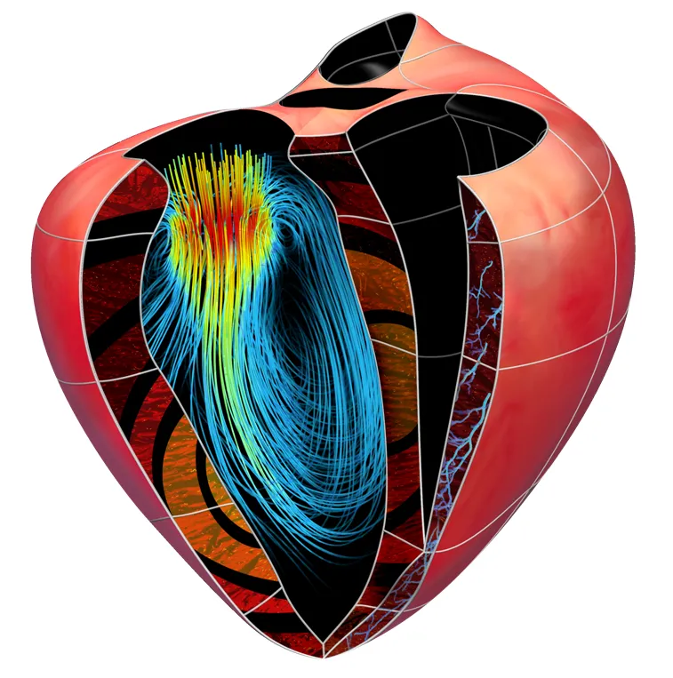
Researchers at King’s have pioneered a method using a 3D printer to create a physical replica of a patient’s organ, to help surgeons plan complex procedures. This is particularly useful for operating on young children, whose organs are very small.
Two-year old Mina was one of the first patients in the UK to benefit from this new technique. From birth, her heart was deformed by a large hole between the two chambers. Researchers at King’s used computer software to stitch together more than 120 images of Mina’s heart, creating a 3D image that could be viewed from any angle. Turning this image into a replica was the next step.
With the replica of Mina’s heart printed in plastic, doctors treating her at Evelina London Children’s Hospital were able to see the size and position of the hole and to successfully design a patch for it.
More recently, the method was used to help successfully transplant an adult kidney into a child for the first time.
Two-year-old Lucy suffered heart and kidney failure as a baby. Having undergone surgery for her heart condition, she faced a lifetime of dialysis but was then given a transplant at Great Ormond Street Hospital using a kidney donated by her father, Chris, during a procedure at Guy’s Hospital.
Models of Chris’ kidney and Lucy’s abdomen were produced using a 3D printer so that the surgeons could plan the highly complex operation and rehearse each step with the 3D models.
Lucy has given the model of her stomach and the model of her father’s kidney to the Science Museum. The models are due to go on permanent display at the museum’s Medicine Galleries, opening in 2019.
Speaking to Evening Standard, Mr Pankaj Chandak, a transplant Registrar at Guy’s and St Thomas’ Hospital and a researcher at King’s whose idea it was to use 3D printouts for Lucy’s operation, said: ‘It’s wonderful to see how well Lucy is doing and it’s an honour to know that millions of people of all ages will be able to learn about the models and Lucy’s surgery when they visit the Science Museum.’
To find out more about Guy’s and St Thomas’ Hospital see here.
For more information about kidney dialysis see here.
To read about the use of 3D printed organs, click here and to watch a video about Pankaj’s work, click here.

