These features make ultrasonic tracking applicable in a range of different clinical fields including fetal medicine, cardiology, anaesthesia and pain management to improve procedure safety and efficiency.
Dr Wenfeng Xia
14 May 2020
Ultrasonic Tracking: Novel technology identifies tip of surgical needle during surgeries
New technology from The School of Biomedical Engineering & Imaging Sciences provides surgeons with greater visibility and accuracy for procedures
Researchers at The School of Biomedical Engineering & Imaging Sciences, UCL and UCH have developed a novel surgical needle tracking technology which for the first time identifies the tip of a needle during surgeries.
The project is part of the seven-year research grant Guided Instrumentation for Fetal Therapy and Surgery (GIFT-Surg) also in collaboration with UZ Leuven and KU Leuven, which develops new surgical technology aiding in the treatment of congenital problems in the womb such as twin to twin transfusion syndrome and spina bifida.
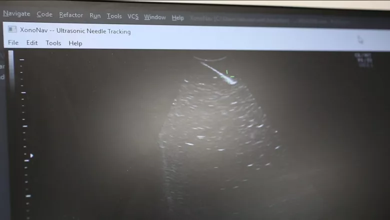
With ultrasonic tracking, a miniature fibre-optic ultrasound sensor is integrated within a thin needle to communicate with an external ultrasound imaging probe.
Dzhoshkun Shakir, Research Software Engineer, who leads the software team of GIFT-Surg, said in ultrasonic needle tracking the aim is to track the tip of a surgical tool such as a needle.
“Our group has developed a new sensor and the software that’s going to operate this technology,” Dr Shakir said.
“We have been developing the software together with hardware engineers who have assembled the ultra-sonic needle tracking console.”
The system, he explained, acquires data from devices such as the GE Healthcare VolusonTM in real time to do advanced processing, to locate the sensor and subsequently visualise the sensor in a live ultrasound video feed from a clinical ultrasound imaging system.
It allows the clinician to clearly see the needle tip when its ultrasound visibility is low. It also prevents the clinicians from potential confusion between the shaft and the tip of the needle, enhancing the accuracy of the needle’s target during procedures.
Wenfeng Xia, co-investigator leading on the ultrasonic tracking project, said ultrasound imaging is widely used for guiding minimally invasive procedures.
“However, these procedures are sometimes challenging as a clinician holds an ultrasound probe which is on the belly of the mother and with the other hand, holds the needle,” he said.
“The two must be aligned so the clinician can see the needle in the ultrasound image. If things are slightly misaligned, it’s easy to confuse the tip of the needle with its shaft.”
The fibre-optic ultrasound sensor has several distinct advantages: it is readily miniaturised so that it can be integrated into a range of different medical devices.
Its broad acoustic bandwidth makes it compatible with various ultrasound probes, and its immunity to an electromagnetic environment allows it to be potentially useful for intraoperative MRI guided surgeries.
With promising results obtained in pre-clinical studies, the team is now preparing for a first-in-human study in 2021 in collaboration with researchers from UCL, and UCLH to provide the clinicians with new information during the surgery to better guide them and enhance patient’s safety.
This project is funded by Wellcome and EPSRC.