Correlative Light and Electron Microscopy

Electron microscopy (EM), although pivotal in life-science research, is limited in its capacity to work with live cells or at the length scales of whole complex tissues. Light microscopy techniques are often employed to image dynamic events in living cells and tissue and/or image complex tissues.

Each technology has its strengths and weaknesses and it is common to use both techniques to investigate cellular systems and events. This is normally undertaken as two separate studies and performed on different samples prepared specifically for light or electron microscopy. Using the same tissue allows an event observed by light microscopy to be directly linked to the higher resolution information provided by EM.

Thus, the capacity of EM is enhanced by correlative light and EM (CLEM) workflows where light microscopy events are co-localised with underlying ultrastructure.

Examples of use

Immuno-electron microscopy is powerful when employed to localise molecules within their functional intracellular locations. However, for rare events or those associated with a dynamic process, it is difficult to have confidence that the structure of interest can be found in a transmission electron microscope. To achieve this we need a specific area to be correlated across length and volume scales. In its most simple form CLEM allows a fluorescent protein, followed by live-cell imaging, to be tracked and processed for imaging by immuno-EM.

We also combine infrared branding in the Nikon Imaging Centre with serial block face scanning electron microscopy to correlate underlying ultrastructure in 3D with electrophysiology studies.

Equipment available

  • JEOL JEM 1400Plus
  • JEOL JEM F200. 
Cell image
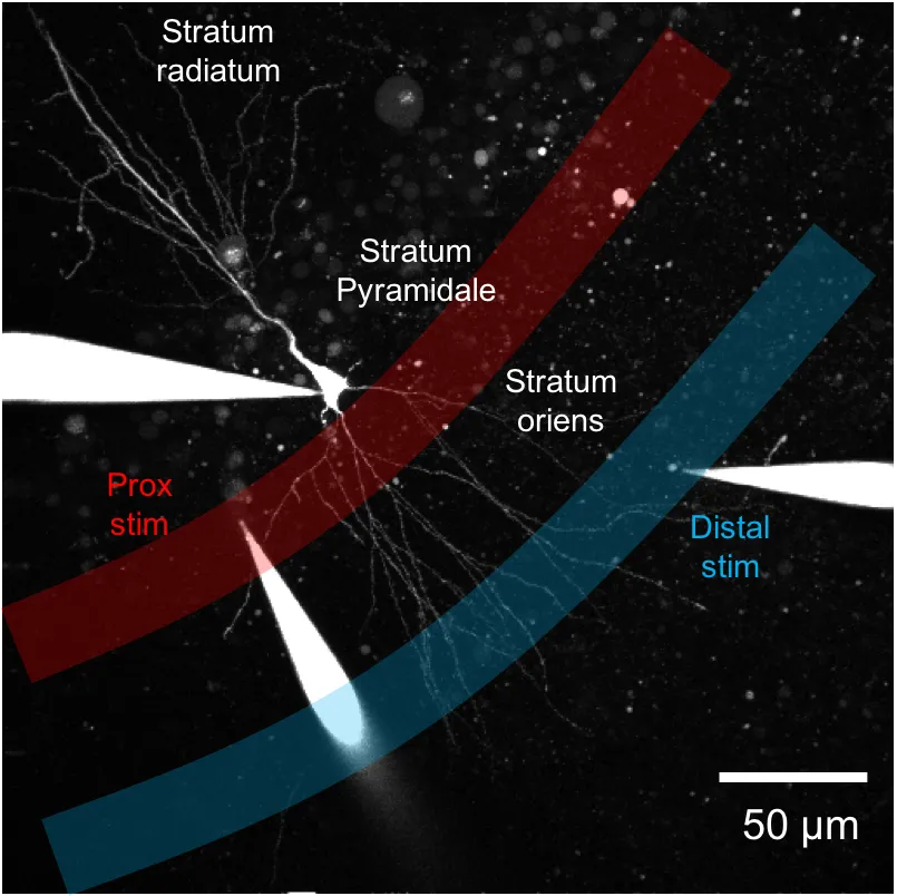
(Electrophysiology investigation of neuronal signaling) To link the electrophysiological response to the underlying ultrastructure not visible by light microscopy it is necessary to perform 3D electron microscopy on the exact same neuron used for the electrophysiology study. This requires a specialised correlative workflow allowing the point of interest to be tracked during tissue processing and to be relocated in x-y-z in the electron microscope.
Cell image
(Infrared branding of tissue) The tissue is branded with an infrared laser to allow the cells to be tracked and located in the electron microscope after processing.
Cell image
The proximal and distal locations previously selected for electrophysiology The proximal and distal locations previously selected for electrophysiology are identified in the serial block face scanning electron microscope. Once located and aligned the microscope collects a stack of 2D images through the areas identified. These image stacks are then reconstructed to generate 3D information relating to the underlying ultrastructure.