Critical Point Drying
Critical point drying (CPD) is a highly effective method to preserve ultrastructure and provide a room temperature stable sample for imaging in a scanning electron microscope.
The sample itself is also normally conductive (due to chemical processing prior to drying) which further aids high-resolution imaging.
The ‘critical point’ is where the physical characteristics of liquid and gaseous states are not distinguishable. Compounds which are at this critical point can be converted into the liquid or gaseous phase without crossing the interfaces between liquid and gas and thus avoiding the damaging effects of the forces of surface tension.
It is important to consider the risk of some air drying of a specimen during the chemical fixation steps and the CPD loading steps.
Example of use
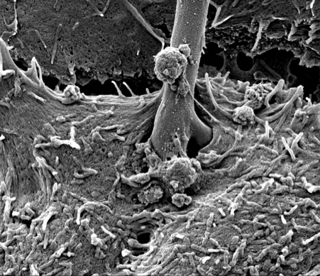
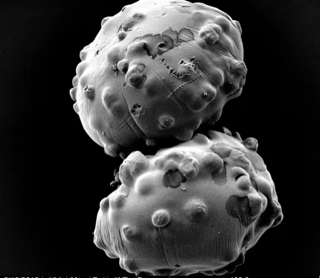
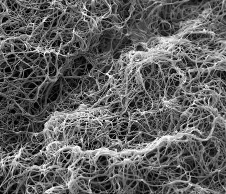
We routinely use CPD to minimise drying artefacts of samples prepared for scanning electron microscopy. For example, when preparing monolayers, tissue and whole organisms such as zebrafish embryos.
Equipment available
- Leica-microsystems EM CPD300


