Cryogenic Transmission Electron Microscopy (Cryo-TEM)
Cryogenic transmission electron microscopy (cryo TEM) can solve near-atomic-resolution structures with volume and clarity.
We have a specially optimised microscope allowing the sample to be studied close to its living state by preserving it through vitrification. We can also operate the microscope at higher accelerating voltages (200kV) and use a field emission gun (FEG) to improve resolutions.
We have also added novel tools to further improve resolution such as:
- an energy filter to improve image contrast and exclude the blurred background. This allows specimens to be imaged closer to focus
- phase plates to give a five-fold increase in contrast
- direct electron detection camera to give unmatched sensitivity and resolution with superior detective quantum efficiency (DQE) than conventional digital cameras. This camera allows movement caused by the electron beam to be corrected computationally and higher resolutions achieved
When combined, these tools offer a paradigm shift in the capability of the microscope and drive advances in cryogenic electron mircroscopy research.
Examples of use
This technique is excellent for:
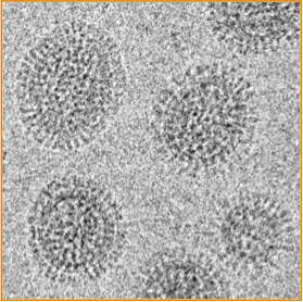
- single particle imaging of protein complexes, viruses, protein-ribonucleic acid associations to understand the fundamental building blocks of life
- structural analysis of membranes in their native and cellular context in 3D at the nanometer-scale
- drug delivery studies (eg colloidal dispersions)
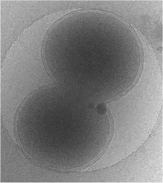
- biotechnology or tissue engineering including characterising synthetic lipid bilayers as models of cell membranes
- studying samples such as lung surfactant, which need to remain as close as possible to liquid phase so its structure and function can be correlated accurately
At the centre we have used cryo TEM to study:
- Plasmodium falciparum erythrocytes during invasion, infection and egress
- antibodies and allergic reactions
Equipment available
- JEOL JEM 2200FS
Images


