Energy Dispersive X-ray Spectroscopy
Energy dispersive x-ray spectroscopy (EDS) (sometimes called energy dispersive x-ray analysis) is an analytical technique used for the elemental analysis or chemical characterisation of a sample.
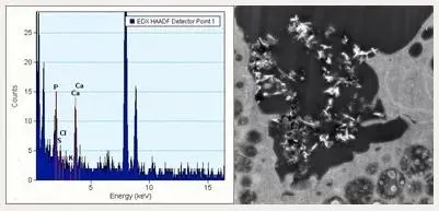
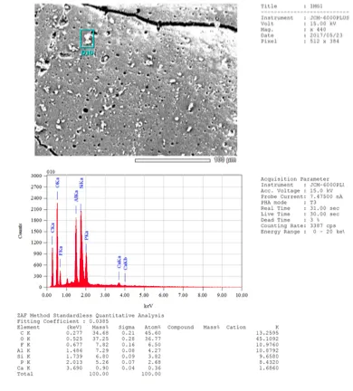
In this technique, when the electron beam is focused onto the area of interest, interactions of the electrons with individual atoms in the specimen result in the generation of x-rays. The collection of these by an EDS detector results in a spectrum that gives both qualitative and quantitative information about the element composition of the specimen.
The characterisation capabilities of EDS are due in large part to the fundamental principle that each element has a unique atomic structure allowing a unique set of peaks on its emission spectrum. Identification of these peaks allows elemental characterisation of a sample.
Examples of use
This technique is useful for:
- routine diagnostic analysis
- environmental pollution studies
- physiology and pathophysiology
- analysis of fluids
- nanotechnology and nanomedicine
When applied to transmission electron microscopy samples, we can use resin sections (eg cell monolayers) or cells that have been grown directly on electron microscopy grids, nanoparticles etc.
When applied to scanning electron microscopy samples, we can use bulk specimens (organic or inorganic), semithin sections etc.
Equipment available
- JEOL JEM F200 TEM
- JEOL JSM 7800 Prime SEM
- JEOL NeoScope JCM 6000Plus.


