Scanning Transmission Electron Microscopy (STEM)
A scanning transmission electron microscope is a conventional transmission electron microscope equipped with additional scanning coils, detectors and necessary circuitry.
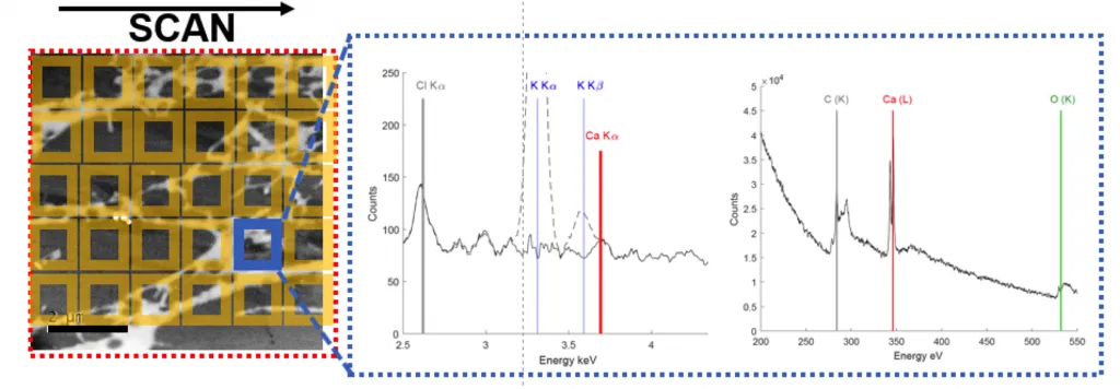
As with conventional transmission electron microscopy (TEM), images are formed by electrons passing through a sufficiently thin specimen, however, in scanning transmission electron microscopy (STEM) the electron beam is focused to a fine spot which is then scanned over the sample in a raster. This is similar to how scanning electron microscopy works. By rastering the beam across the sample, STEM is suitable for analytical techniques such as energy dispersive spectroscopy.
STEM offers a number of potential advantages over TEM imaging for tomography of biological specimens. A practical advantage of STEM is its capacity to generate contrast without the need for defocusing. The scanning geometry of STEM also allows for dynamic focusing so that the imaging conditions remain uniform across a tilted specimen. STEM can allow tomography of thicker sections with improved signal-to-noise ratio and contrast over conventional TEM tomography. This has significant potential for understanding subcellular organisation and organelle-organelle interactions at the nanometre scale through a whole cell volume.
STEM tomography is a new and emerging technique in life science research. We are collaborating with leading international groups to advance this technique and understand its full potential.
Examples of use
This technique is useful for:
- antitumoral therapies (eg platinum based drugs, SPIONs etc)
- cell viability studies and nanotoxicology
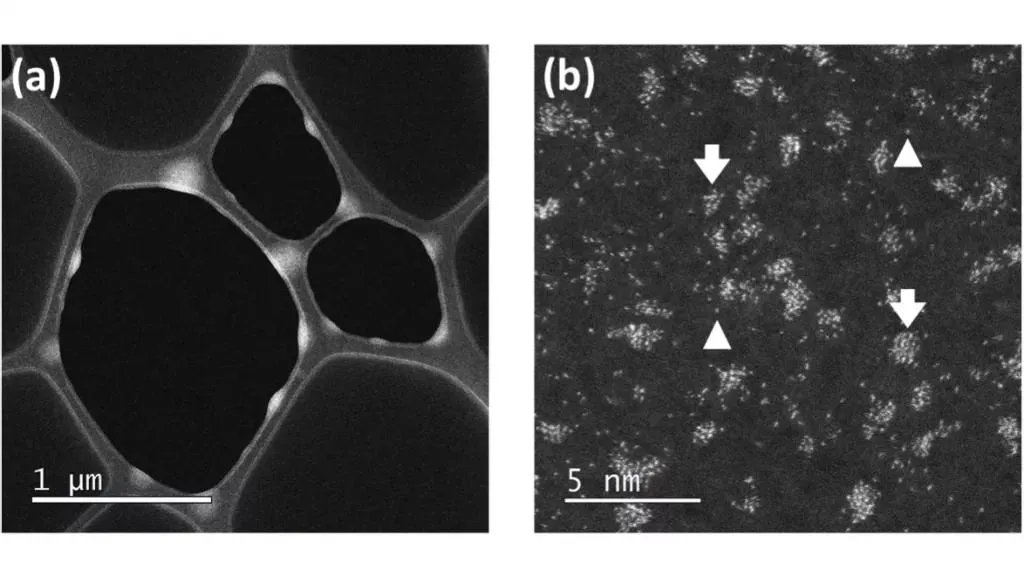
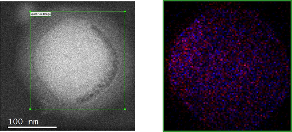
An example of this technique is the localisation of nanoparticle uptake into cells. STEM is most often employed for physics and chemistry studies. In these studies, z-contrast annular dark-field imaging and spectroscopic mapping by energy dispersive spectroscopy are used. The highly focused beam scatters electrons depending upon the atomic number of atoms in the specimen. Areas of high brightness indicate the presence of high atomic number elements in the specimen.
Equipment available
- JEOL JEM F200.


