Tokuyasu Technique and Immunogold Labelling
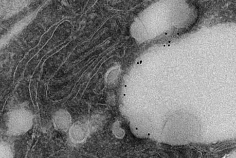
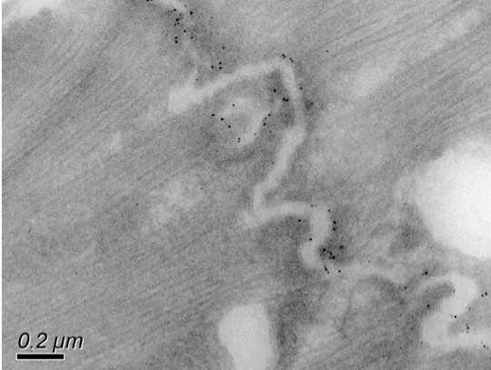
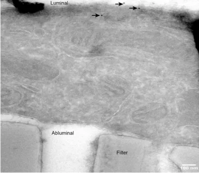
Tokuyasu cryosectioning technique allows immune labelling and therefore the detection of specific proteins in cells by electron microscopy. By conjugating a sample specific antibody to a small colloidal gold particle (eg 1nm, 5nm, 10nm, 20nm) labelled structures can be identified using the a transmission electron microscope.
Tokuyasu technique generally surpasses other techniques in membrane visibility and labelling sensitivity. The method follows a simple protocol: cells are lightly chemically fixed, after which they are frozen in the presence of sucrose and cut into 50-100 nm cryosections. The sections are then thawed and labeled with antibodies that specifically recognize the molecule of interest, while coupled to gold particles that are detectable in the microscope.
It complements fluorescent labelling by localising immune reactions to specific intracellular structures at a resolution that cannot be achieved with fluorescence. For this reason it has become a powerful tool when it comes to prove co-localisation of antigens. The technique is also used at scanning electron microscope level to provide details about the surface distribution of antigens.
Examples of use
This technique is useful for studying:
- intracellular localisation of molecules
- intracellular relationships between molecules (protein co-localisation)
We routinely use this technique to:
- study the localisation of synaptic proteins associated to synaptic dysfunction at the neuromuscular junction in ALS disease
- investigate the role for Nup98 in virus morphogenesis
Equipment available
- Leica UC7 cryo-ultramicrotome
- JEOL JEM 1400Plus
- JEOL JEM F200.


