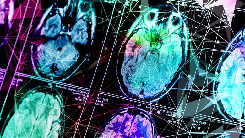
Our physicists, principally led by Professor Jo Hajnal, have rebuilt the way imaging is done for babies for the last 20-30 years, and we can now get very high-quality imaging of newborn babies.
Professor David Edwards, consultant neonatologist at the Evelina London Children's Hospital and Professor of Pediatrics & Neonatal Medicine at King's College London
06 April 2022
Brain charts map the rapid growth and slow decline of the human brain over our lifetime
The dHCP, a large open science project is an important part of the paper, having provided high quality, otherwise difficult to obtain brain MRIs of newborn babies
Researchers from the Developing Human Connectome Project (dHCP) were part of an international team of researchers that created a series of brain charts spanning our entire lifespan – from a 15-week-old fetus to 100-year-old adult – that show how our brains expand rapidly in early life and slowly shrink as we age.
The charts are the result of a research project spanning six continents and bringing together possibly the largest ever MRI datasets ever aggregated – almost 125,000 brain scans from over 100 different studies. Although not currently intended for clinical use, the team hopes the charts will become a routine clinical tool similar to how standardised paediatric growth charts are used.
Growth charts have been a cornerstone of paediatric healthcare for over 200 years and are used ubiquitously in clinics to help monitor the growth and development of children in comparison to their peers. A typical growth chart might plot age on the horizontal axis versus height on the vertical axis, but rather than being a single line, it will show a range that reflects the natural variability in height, weight or head circumference.
There are no analogous reference charts for measuring age-related changes in the human brain. The lack of tools for standardised assessment of brain development and aging is particularly relevant to the study of psychiatric disorders, where the differences between conditions and the heterogeneity within them demands instruments that can say something meaningful about a single individual in the way clinical reference charts can, and to conditions such as Alzheimer’s disease that cause degeneration of brain tissue and cognitive decline.
Today’s study, published in Nature, is a major step towards filling this gap. Unlike paediatric growth charts, BrainChart – published on the open access site brainchart.io – covers the whole lifespan, from development in the womb through to old age, and aims to create a common language to describe the variability in brain development and maturation.
The incredible growing and shrinking brain
The brain charts have allowed the researchers to confirm – and in some cases, show for the first time – developmental milestones that have previously only been hypothesised, such as at what age the brain’s major tissue classes reach peak volume and when do specific regions of the brain reach maturity.
The dHCP, a large open science project is an important part of the paper, having provided high quality, otherwise difficult to obtain brain MRIs of newborn babies.
Professor David Edwards, consultant neonatologist at the Evelina London Children's Hospital and Professor of Paediatrics & Neonatal Medicine at King's College London said obviously the brain of a baby needs detailed imaging – an ordinary scanner cannot be used nor sequences that are used for an adult.
The scans which take about an hour are multimodal and both structural and functional led the team to uncover a part of the brain, the insular, which is an interesting part of the brain which keeps growing far longer than was understood until researchers saw the data.
The data helps to unpick how the brain develops and understand how that might or might not go wrong. The dHCP researchers are not only looking at the whole brain, but also the individual sections and how they do or do not grow together. It is in this way that it will enable researchers to understand more how the brain has evolved and eventually how it is functioning.
“The dHCP data is open source and this type of work is a particularly good use of the data. We hope that more researchers will be inclined to use it.”
Dr Richard Bethlehem from the Department of Psychiatry at the University of Cambridge, one of the co-leads of the study, said: “One of the things we’ve been able to do, through a very concerted global effort, is to stitch together data across the whole life span. It’s allowed us to measure the very early, rapid changes that are happening in the brain, and the long, slow decline as we age.”
Among the key milestones observed by the team were:
- The volume of grey matter (brain cells) increases rapidly from mid-gestation onwards, peaking just before we are six years old. It then begins to decrease slowly.
- The volume of white matter (brain connections) also increased rapidly from mid-gestation through early childhood and peaks just before we are 29 years old.
- The decline in white matter volume begins to accelerate after 50 years.
- Grey matter volume in the subcortex (which controls bodily functions and basic behaviour) peaks in adolescence at 14-and-a-half years old.
The team have created the tool with a reference framework to allow other researchers and clinicians to adjust their own datasets, making it possible to compare them against the BrainChart population.
The NHS does millions of brain scans every year and in most of these cases, they are assessed by radiologists or neurologists relying on their extensive expertise to judge whether there is anything clinically relevant apparent on these scans. We hope that clinicians in future will be able to compare their data against ours and produce a more comprehensive report adding additional objective and quantitative observations to their assessment.
Dr Richard Bethlehem from the Department of Psychiatry at the University of Cambridge
“This should effectively allow the neurologist to answer the question ‘this area looks atypical but atypical by how much?’. As the tool is standardised, it shouldn’t matter where you have your brain scan – you should still be able to compare it.”
The research was supported by the British Academy, the Autism Centre of Excellence, the Medical Research Council, National Institute for Health Research (NIHR), the Wellcome Trust and the NIHR Cambridge Biomedical Research Centre.
In this story

Professor of Imaging Science

Director, Insititute for Women & Children's Health