The MAGNETOM Free.Max MRI scanner is a prime example of how our School effectively works with industry to engineer better health. Together with Siemens Healthineers, we are ultimately creating more healthcare opportunities for clinicians equipping them with technology necessary to better deliver patient care, not only in standard hospital settings, but also within resource limited areas in the broader community. The key funding from Research England, which allowed for the enabling works for the scanner to be fitted, bolsters the strategic objective of St Thomas’ MedTech Hub and the London Institute for Healthcare Engineering.
Professor Sebastien Ourselin, Head of the School of Biomedical Engineering & Imaging Sciences at King’s College London
17 August 2022
First in the UK: new MRI scanner enables use outside of hospital setting, accelerating new research opportunities
King’s College London leads research into breaking the barriers of conventional MRI with the installation of the UK’s first MAGNETOM Free.Max.
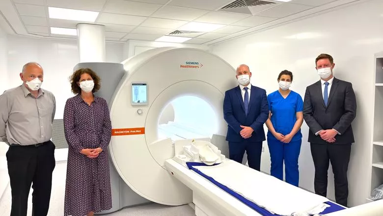
King’s College London (King’s) is driving efforts to make new MRI technology more accessible in community settings through virtually helium-free MRI research with the first MAGNETOM Free.Max in the UK from Siemens Healthineers. The reduced-helium MRI allows the King’s team to evaluate this type of MRI for use outside of a traditional hospital setting, while also using a lower field strength, expanding the scope of research into cardiac, respiratory, and foetal brain development imaging that was previously not possible.
The delivery of the first MAGNETOM Free.Max at King’s College London, alongside funding by Research England, breaks the barriers of conventional MRI, enabling research previously not possible for the department.
Unlike conventional MRI systems, the MAGNETOM Free.Max requires less than one litre of liquid helium for cooling, an increasingly scarce resource, eliminating the need for a quench pipe which is otherwise used to safely and quickly expel helium out of a scanner in case of an emergency. The virtually helium-free MRI scanner radically reduces infrastructure requirements, enabling it to be installed in community-based settings.
King’s College London, one of the world leading universities in imaging sciences, has an existing relationship with Siemens Healthineers and has partnered with the Research and Development team to provide additional insight into research projects utilising other MRI systems including 1.5T, 3T and 7T MRI systems from Siemens Healthineers.
The addition of the 0.55T MAGNETOM Free.Max within the King’s Advanced MRI Centre at St Thomas’ Hospital (part of Guy’s and St Thomas’ NHS Foundation Trust) expands research capabilities and exploits currently existing high-tech facilities, such as a Radio Frequency Coil Lab.
The research team at King’s College London plans to use the lower field of 0.55T for imaging research into areas that are more challenging for MRI at 1.5T and above, such as respiratory imaging, interventional radiology, and scanning patients with implants as diagnostic capabilities are strongly improved.
This first of its kind MRI also features an 80cm bore, the entry space for patients, which will provide a more comfortable scanning experience for claustrophobic and bariatric patients, increasing the likelihood of participation in studies. Augmented with AI, the system supports the automation of routine examinations – reducing cognitive load for researchers and opening new possibilities for radiographers faced with increasing time pressures.
The virtually helium-free infrastructure of the MAGNETOM Free.Max helps to break the barriers of siting constraints and provides possibilities to expand the reach of MRI not previously possible with conventional MRI systems. We are pleased to be partnering with the team at King’s College London to maximise research opportunities into exploring resolutions for repeat MRI cases and helping to emulate a community diagnostic centre setup within the Advanced Imaging Centre.
Dr Craig Buckley, Head of Research and Scientific Collaborations at Siemens Healthineers GB&I
The MAGNETOM Free.Max from Siemens Healthineers is a welcome addition to King’s College London as we seek to evaluate how this kind of MRI might perform outside of a hospital setting, and expand the scope of our imaging research with a lower field strength. Community diagnostic centres are the setting we’re trying to simulate here with the MAGNETOM Free.Max. Whilst the system is currently used in a hospital setting, its less demanding infrastructure requirements means it could be implemented into community settings, increasing MRI accessibility for patients.
Dr Sharon Giles, Director of Clinical & Research Imaging Operations, School of Biomedical Engineering & Imaging Sciences at King’s College London
In this story

Assistant Principal (Innovation)
Director of Clinical & Research Imaging Operations