The 7T scanner – while very powerful – has not previously been cleared for use in infants. We performed very detailed safety tests in-house and modified the scanner’s software to add a margin for safety. This process included working with industry and conducting scientific studies of how infants may experience being scanned. By working in this cross-disciplinary way, we have created an almost unique capability in the world that we are immensely proud of, and are very excited to see where this takes us.
Dr Shaihan Malik, Head of the Department of Imaging Physics & Engineering, School of Biomedical Engineering & Imaging Sciences
01 March 2024
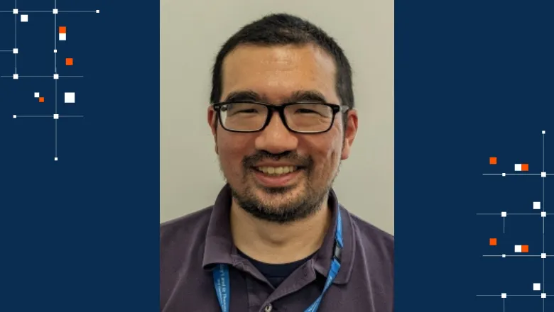
In Conversation with Dr Tomoki Arichi
Dr Tomoki Arichi is the Head of the Department of Early Life Imaging at the School, and an Honourary Consultant at the Evelina London Children's Hospital. We spoke to Tomoki about how his clinical experience drew him to research in paediatric neurodisability, and how academic and clinical collaboration can make groundbreaking research possible.
Dr Arichi has been working with the School since 2012, after completing his PhD at Imperial College London. His research is centred around developing and applying novel imaging methods for studying the developing human brain. "A particular focus is understanding how activity first emerges in the brain, which I study using functional MRI methods," said Dr Arichi.
When asked why he was drawn to this line of study, he explained, "In my clinical work, I manage the care of children who unfortunately have long-term neurodevelopmental difficulties as a result of a brain injury around the time of birth. Through this work, I realised there is so much we don’t know about how the brain first develops, and how this development is influenced by diseases and environmental factors. Unfortunately, we can only provide treatments for symptoms when they arise, as we don’t have the mechanistic insight to definitively cure these problems."
Recently, Dr Arichi was awarded a £2.1 million Senior Clinical Fellowship grant from the Medical Research Council (MRC). The funding will enable a new programme of cutting-edge research which he hopes will markedly increase our understanding of how the human brain first develops.
"The Fellowship will study how the outermost layer of the brain (the cortex) develops in the crucial period around birth. Activity in the cortex is essential to allow interaction with the world around us and with other people. However, it is not known how the cortex and its activity first mature at the start of our lives, or how the cortex changes to allow young children to develop learning and socialising skills as toddlers. The goal of my research is to learn how abnormal cortical development can arise in early human life, and how it can lead to difficulties with behaviour and learning in later childhood," says Dr Arichi.
Supporting his research will be the ultra-high field 7 Tesla MRI scanner recently installed at the School's imaging facilities in St Thomas' Hospital. "We are extremely privileged to have one of the few 7T MRI scanners installed in a hospital setting in the entire world, meaning we are uniquely placed to study this subject in detail, especially in terms of imaging very young and fragile newborn infants. This work is completely unique worldwide and due to the much higher detail provided by a 7T scanner, we can gain entirely new insights into brain activity in this crucial period of early life."
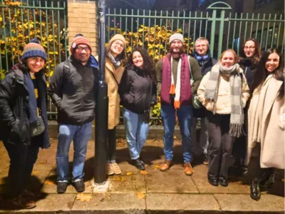
Dr Arichi believes that pioneering research is made possible by the diverse teams that come together to deliver it. "I feel very fortunate to be working at King’s and in the School, as the combination of the clinical setting with the unparalleled imaging facilities means that we have unique opportunities to perform cutting-edge studies that can have a real impact on patients. Key to all of this are the incredible colleagues that I work with, who all have synergistic but diverse expertise. The 7T work, for instance, was only made possible by working closely with Dr Shaihan Malik and Pip Bridgen, as well as a whole team of incredible PhD students and neuroscientists.
Scanning of Neonates has been an amazing achievement for everyone involved. It was a privilege to work on this with Tom and develop the sequences used. There were many challenges to overcome due to the novelty of the project, but our ability to now scan neonates at 7T has the potential to give improved diagnoses, gain new information about pathophysiology, and provide novel insights into early brain development. Overall, this can help inform treatment plans for the infants that are scanned, thereby allowing us to offer improved patient care for them.
Pip Bridgen, Superintendent Advanced MRI Research Radiographer, St Thomas' Hospital
Dr Arichi will be studying a group of toddlers to understand the brain changes underlying new behaviours as they emerge in early childhood: "Studying these phenomena in detail was previously not possible - partly because it is extremely challenging to study young children inside an MRI scanner. We have developed a new virtual reality (VR) system that provides a fun and immersive experience for a child having an MRI scan; for the first time, it can allow detailed pictures of the brain to be acquired from a child whilst they are awake and playing in the VR environment."
Dr Arichi hopes that the impact of this research will go beyond academia at King's. "The results will provide unprecedented knowledge about early cortical development and its effects on early interaction in humans. I look forward to sharing this information and methods openly so that they can be used by the scientific community to answer fundamental questions about the cortex and its development."
This new knowledge will help doctors and scientists identify which children may have difficulties later in life, devise and start new treatments to prevent or treat abnormalities in their cortex and help to understand how they may cause conditions such as learning difficulties, autism spectrum conditions, and mental health disorders. Ultimately, I really hope that my research can change things for the better for vulnerable children and families.
Dr Tomoki Arichi, Head of the Department of Early Life Imaging and Clinical Reader in Paediatric Neurodisability, School of Biomedical Engineering & Imaging Sciences
In this story
Head of the Department of Early Life Imaging
Head of the Department of Imaging Physics and Engineering
Superintendent Advanced MRI Research Radiographer