This project shows how far we have advanced technologically, while maintaining the desire for preservation. While we could have scanned the entire skeleton and replaced the original completely,, restoring it in this way allows for a greater appreciation of the history of this skeleton. It at least partially retains its authenticity and widens the path for more restoration projects like this to preserve fragile specimens that would otherwise be unavailable to view.
Asma Bint-Zubair, the Royal Veterinary College
17 April 2023
Medical 3D printing restores 100 year old Museum of Life Sciences specimen
Prof Kawal Rhode, School of Biomedical Engineering & Imaging Sciences, and Royal Veterinary College Student, Asma Bint-Zubair, have restored a 100 year old chimpanzee skeleton using CT scans and novel 3D printing techniques.
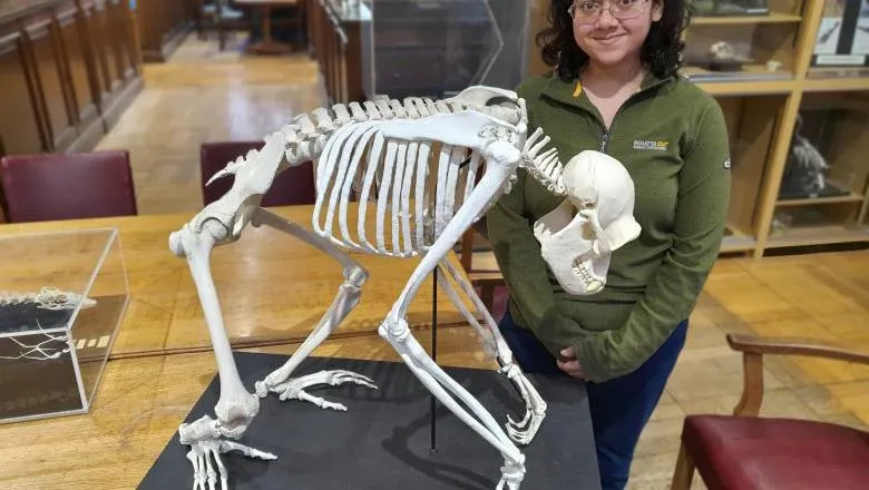
The chimpanzee skeleton was originally acquired by Prof Arthur Dendy who was appointed Professor of Zoology at King's in 1905.
The exact age of the skeleton is unknown, but at over 100 years old the damage would have occurred over time since real dry bones are brittle and very fragile. The skeleton was transported to its current location in the Hodgkin Building at Guy's from the Strand Campus over 20 years ago where repair efforts were made, but already a large part of it was missing and it has remained in this condition until now.
Museum of Life Sciences Curator, Dr Gillian Sales, explains:
"In the Museum we use the chimp skeleton for teaching about primates and the changes that occur in posture and locomotion during the emergence of man. The badly damaged skeleton was of little use in demonstrating the anatomical features highlighted in these sessions. When it became clear that modern technology could perhaps remedy this, I approached Prof Rhode to see if it would be possible to do so.
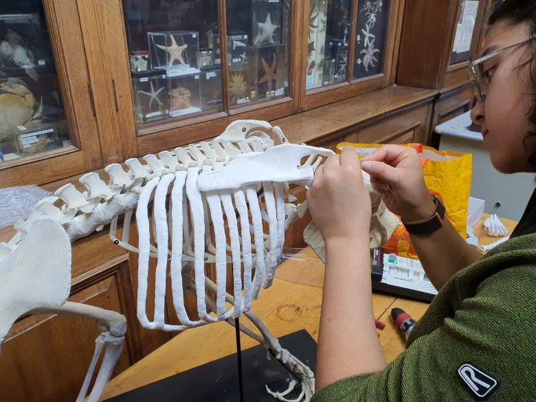
"Many of the specimens were originally prepared many years ago, some over 100 years ago, but people showed amazing skill in preparing skeletons, e.g. wiring small bones together, replicating the form that the animal would have had in the wild, preserving plants so that they can be appreciated today often in their original colour. A similar level of skill has been used in this project in the scanning process to get the appropriate data for manipulation, in the printing process and especially in the final treatment of the printed parts to remove surplus material while preserving essential features."
This is a really interesting case where we developed and learnt the techniques from the clinical world and then translated these to the animal world. We have been working with the thoracic surgeons at Guy's hospital to rebuild the rib cages of lung cancer patients whose bones have been invaded by cancer. We take a CT scan of the patient and then use this to 3D print the affected bones. We used exactly these techniques to rebuild the skeleton of our chimpanzee.
Prof Kawal Rhode, Head of Education, School of Biomedical Engineering & Imaging Sciences
Future collaborative plans include looking at the Museum collection to identify other skeletons that could benefit from the techniques utilised in this restoration.
3D printing techniques can also be used to create replicas that visitors and students can handle without fear of damage.
The medical applications of the 3D printing techniques featured in this story were recently covered by the BBC on their science programme, Click.
The Museum of Life Sciences exists to celebrate and explain the diversity of animal and plant life in the context of the biological and health sciences. Opened in 2009, it brings together historic biological and pharmaceutical collections from the constituent colleges that make up the modern College.
In this story

Professor in Biomedical Engineering and the Head of Education at the School of Biomedical Engineering & Imaging Sciences