This work is important as a promising COVID diagnostic and prognostic biomarker has been developed but needs to be tested prospectively in different centres across the UK where there are very different populations.
Dr Tom Booth, Senior Lecturer in Neuroimaging
18 January 2021
Neuroradiology research team receive funding to develop COVID-19 biomarker
The research has huge implications for patient ward allocation and patient management.

A team of researchers from King’s College Hospital (KCH), King’s College London School of Biomedical Engineering & Imaging Sciences and Addenbrooke’s Hospital in Cambridge have been awarded with a research grant from Radiological Research Trust, an NIHR partner and charity which provides funding for research and education in medical imaging, to carry out research into the use of neuroradiology techniques in the COVID-19 diagnosis.
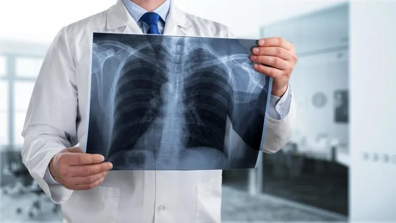
In September last year, a team from the School of Biomedical Engineering & Imaging Sciences led by Dr Tom Booth, senior lecturer in neuroradiology at King's College London and consultant neuroradiologist at KCH, identified a biomarker that could identify COVID-19 infection from routine emergency chest scans used to diagnose stroke.
The team now aim to collect patient data from hospitals across the UK in order to further develop and validate this biomarker as an effective tool in the rapid diagnosis of COVID-19.
Chief Investigator Dr Tom Booth will oversee the study and it will be led by KCH neuroradiology fellow Dr Nathan Chan alongside Dr Tarini Ratneswaren, neuroradiology fellow at Addenbrooke’s Hospital, Cambridge.
“If the biomarker is shown to work accurately in different scenarios throughout the UK, we can conclude that the biomarker has been fully developed and validated.”
“The biomarker is free information obtained opportunistically from a routine urgent stroke scan.”
“The biomarker is available very early on in the patient pathway well before SARS COV-2 PCR test results are ready. This has huge implications for patient ward allocation and patient management.”
Dr Chan said the validation of the COVID-19 imaging biomarker will contribute to faster diagnosis and better management of acute stroke patients.
On behalf of the team working on the COVID-19 stroke apical lung examination study and the radiology academic network for trainees (RADIANT), we would like to thank the Radiological Research Trust for its most generous contribution to our work.
Dr Nathan Chan
The Radiological Research Trust is committed to supporting new and novel research in medical imaging through the award of small grants to encourage pump priming.
“We particularly welcome grant applications from new and trainee researchers, and over the last year have been keen to do our bit to support COVID-19 efforts,” an RRT spokesperson said.
“The Trustees were impressed with Dr. Chan’s and Dr Booth’s clear and timely proposal, in addition to the proposed team assembled to carry out the research. We are excited to see the impact of the lung biomarker on COVID-19 diagnosis and prognosis, and hope that this work leads to improved clinical outcomes.”
Radiological Research Trust
Dr Ratneswaren said there is initial interest from 46 hospitals, potentially allowing acquisition of a large volume of data with representative data of the whole population of the UK to validate the use of CT of the lung apices as a diagnostic and prognostic biomarker for COVID in stroke patients.
“This is really exciting as it has the potential to impact patient management across the country,” she said.
Dr Chan and Dr Ratneswaren are members of the Radiology Academic Network for Trainees (RADIANT), a network which links trainee radiologists from across the country to deliver research studies and facilitate recruitment to NIHR CRN portfolio studies.

