Sodium imaging has huge potential because it allows us to access critical new measures of brain function. It is exciting because we are now on the cusp of realising this potential owing to the availability of ultra-high field imaging technology.
Dr David Carmichael, Reader in Magnetic Resonance Imaging, School of Biomedical Engineering & Imaging Sciences
05 October 2022
Researchers commence sodium scanning to provide new information for epilepsy diagnosis
Researchers and clinicians have started scanning sodium in the brain to learn more about epilepsy in children
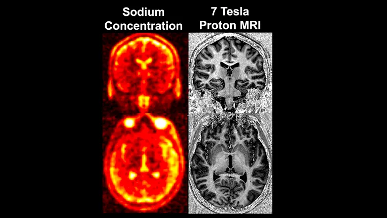
For the first time researchers and clinicians from the School of Biomedical Engineering & Imaging Sciences, UCL, University of Melbourne and Guy’s & St Thomas’ NHS Foundation Trust, have started using Magnetic Resonance Imaging (MRI) to image sodium in the brain to see if it can provide new information to help clinicians detect and understand epilepsy.
Epilepsy is a very common disease where patients have repeated seizures that are due to cascades of unwanted electrical activity (‘firing’) of brain cells. This is often because the brain cells fire too easily.
How easily the ‘firing’ occurs depends on the flow of two elements – sodium and potassium – in and out of the brain cells. In some people there is a genetic error that means sodium flow is abnormal leading to cells firing too easily and generating seizures.
Lead researcher and Reader in Magnetic Resonance Imaging, Dr David Carmichael said in children some of the more severe forms of epilepsy are caused by these alterations in sodium.
Funded by the Wellcome EPSRC Centre for Medical Engineering, the project is using the high field strength 7T scanner in the School’s Advanced MRI Suite, located in St Thomas’ Hospital, to take the images of the rare element, sodium.
The researchers have so far taken images in a small group of 10 children with and without a particular genetic abnormality that causes alterations in brain sodium.
Sodium in the body has an electrical charge, and electrical signals in the brain are created when the sodium travels from the inside to the outside of brain cells.
The MRI signal from sodium within the body is very small, thus making sodium imaging technically very challenging. As a result, this work has involved close collaboration with physicists from King’s College London, the University of Melbourne and UCL to establish the technique on the 7T scanner.
Some patients have genetic abnormalities which can affect the sodium channels that control this process. Mutations in genes encoding sodium channels are the most frequently identified group of genes associated with epilepsy with SCN1A (a gene that belongs to a family of genes that provides instructions for making sodium channels) the largest contributor.
This can lead to uncontrolled electrical signals that can generate seizures in patients with epilepsy and these genetic types of epilepsy are often hard to control with medication and have significant morbidity and mortality.
However, the mechanisms by which these genetic defects cause epilepsy are still not well understood.
The long-term aim of this research is to develop a better understanding and treatment of sodium channel abnormalities that cause epilepsy through direct in-vivo imaging of brain sodium.
We are excited to pilot sodium imaging at 7T in patients with epilepsy caused by loss of function sodium channel mutations. This offers an untapped potential as a non-invasive method to map sodium channel function and correlations with disease severity in these patients. We can also learn more about sodium functional networks which are unknown. As more advanced therapies are being developed for genetic disorders, the need for an objective method to measure impact and response of disease treatments has never been more paramount.
Dr Shan Tang, Consultant Paediatric Neurologist at Evelina London Children’s Hospital
As neuroradiologists, we know we are under diagnosing causes of epilepsy on conventional hospital MRI scans, particularly in children. The higher field strength of 7T MRI, which enables this novel imaging of brain sodium, could be a significant step in providing additional information to improve diagnosis and monitor treatment in epilepsy – ultimately benefitting patients and their parents.
Dr Jon Cleary, Pan-London Neuroradiology Fellow and honorary researcher at the School of Biomedical Engineering & Imaging Sciences
In this story

Professor in Magnetic Resonance Imaging