Brain cancer is uncommon but is disproportionately devastating. The fact that it is uncommon means resources need to be combined worldwide to run imaging studies with sufficient numbers. This work highlights the evidence gaps and provides a focus for combining resources to tackle these gaps.
Dr Tom Booth, Senior Lecturer in Neuroimaging at the School of Biomedical Engineering & Imaging Sciences and Consultant Diagnostic and Interventional Neuroradiologist at King’s College Hospital
04 March 2022
Researchers put forward evidence for use of advanced MRI techniques as brain cancer monitoring biomarkers
The statement is a result of a joint effort of researchers from eight European countries and the US, highlighting evidence gaps and provides a focus for combining resources to tackle these gaps
Researchers from the School of Biomedical Engineering & Imaging Sciences and the Glioma MR Imaging 2.0 (GliMR) initiative, have released a two-part position statement summarising evidence for use of advanced MRI techniques as brain cancer monitoring biomarkers in the clinic, highlighting the latest bench-to-bedside developments. They also discuss emerging topics and indicate the evidence gaps, strengths and limitations. The statement is a result of a joint effort of researchers from eight European countries and the US.
Published in Frontiers in Oncology, the first publication contains imaging relating to brain cancer blood flow as well as structure on a microscopic scale. The second publication contains imaging techniques related to tissue chemical composition as well as the processing of multiple images resulting from different techniques.
The lead on the position statement, Dr Thomas Booth who is a Senior Lecturer in Neuroimaging at the School of Biomedical Engineering & Imaging Sciences and Consultant Diagnostic and Interventional Neuroradiologist at King’s College Hospital, said the researchers sought to make a position statement regarding advanced magnetic resonance imaging in brain cancer that is of sufficient rigour to be the “go-to” reference that remains relevant for the next decade.
“In our position statement, we summarize the current readiness highlighting how the individual techniques are ready or not in terms of technical development and also whether they are ready or not for the clinic.”
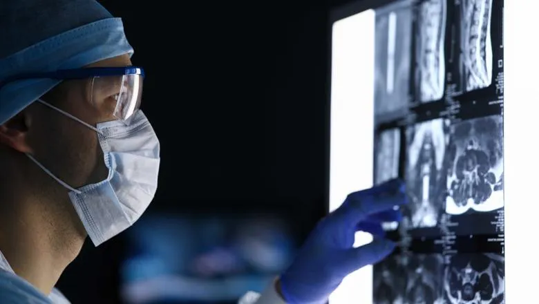
Dr Booth said standard MRI is typically used to monitor brain cancer in the clinic. However, the images are not specific which means that the same MRI appearances could show either the presence of growing tumour or that the tumour has been treated effectively.
“Standard magnetic resonance imaging is not fit for purpose. We cannot tell if someone is responding to treatment or not. That has motivated the proliferation of research into advanced magnetic resonance imaging.”
In the first publication, the researchers show that considerable progress has been made in the development of monitoring biomarkers of blood flow and structure.
Many techniques are still in their infancy such as advanced microstructure imaging techniques, whereas others have generated a larger body of evidence for clinical application like dynamic susceptibilty-weighted MRI, a blood flow imaging technique.
The analysis also highlighted the need for optimal acquisition protocols and mode of processing analysis. Also, the parameter of highest diagnostic value and optimal cut-off points were highlighted as needing to be established.
In the second publication, the researchers summarise advanced MRI techniques that assess chemical composition of tumour tissue and show that these techniques hold huge promise in treatment response assessment.
The researchers’ clinical readiness analysis highlights that most monitoring biomarkers require standardised international consensus guidelines, with more facilitation regarding technique implementation as well as steps to enable seamless reporting in the clinic.
Two comprehensive position statement publications were required to pool the expertise of established, as well as up and coming, leading researchers across Europe and North America. As magnetic resonance imaging works so well in the brain (compared to many other parts of the body), these advanced imaging techniques are typically first finessed in the brain which means there are plenty of state-of-the-art advanced imaging techniques to describe.
Some of the emerging techniques include multi-echo perfusion sequences which make less corrections after imaging necessary. Fractional tumour burden dynamic susceptibilty-weighted MRI maps may translate to the clinic and give a more nuanced interpretation of how tumours respond to treatment. Blood flow measurements such as dynamic contrast-enhanced MRI or arterial spin labelling benefit from higher MRI field strengths via increased signal-to-noise ratios. Moreover, arterial spin labeling allows for blood flow measurements without an injection of contrast agent, which has great potential for clinical use.
Longitudinal methods to determine treatment response, i.e. digitally comparing a new image to an old image, are becoming more established in simple microstructure imaging techniques. More advanced microstructure imaging techniques aim to simultaneously incorporate flow in blood vessels or reflect actual tissue compartments, such as the extra-axonal or extracellular space, intra-cellular, or intra-axonal space, and fluid, such as oedema or cerebrospinal fluid.
Spectroscopic techniques at ultra-high field strength are now better equipped to measure metabolites such as glycine and glutamine. Nuclei other than protons also show promise in spectroscopy or imaging such as phosphorus, deuterium, carbon and sodium. Labile protons on endogenous proteins can be selectively targeted with chemical exchange saturation transfer imaging which is in a state of rapid development.
Multi-modality techniques where different advanced MRI biomarkers are combined with each other or with radionuclides are also promising for determining response to treatment. Other ways of using MRI data once it has been acquired relates to machine learning techniques of augmentation, transfer learning, and techniques to “learn with few examples”. Additionally, federated learning, aka bringing the code to the data, instead of the data to the code, may be a more practical way to help with the problem of having a relatively low incidence of glioma and having few useful datasets.
Dr Booth said the next steps for the research will be for clinicians, engineers, and physicists with expertise in this field to convene through the European Cooperation in Science and Technology (COST) Glioma MR Imaging 2.0 initiative, a pan-European and multidisciplinary network to better inform and enhance development and application of advanced MR imaging.
This group of experts has the opportunity to carry out two actions relating to this work. First, to reiterate, we need to combine resources worldwide to make technical progress and run studies that are of sufficient size to be meaningful. Second, this work provides the foundation for producing a multi-stakeholder downstream guideline for each individual modality. This work highlights the evidence gaps and provides a focus for producing such guidelines as well as pooling resources to tackle these.
Dr Tom Booth, Senior Lecturer in Neuroimaging at the School of Biomedical Engineering & Imaging Sciences and Consultant Diagnostic and Interventional Neuroradiologist at King’s College Hospital
In this story

Reader in Neuroimaging