This finding is the culmination of years of research and technological development at CLF and our partner institutions and we’re extremely excited about its potential to inform the course of cancer research going forward. If this interface proves to be an effective therapeutic target, it could provide an entirely new approach to much needed pharmaceutical development.”
Professor Marisa Martin-Fernandez, Leader of the Octopus Group at CLF, which led the study
28 March 2024
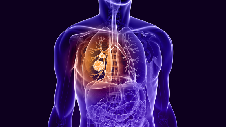
Scientists identify Achilles heel of lung cancer protein
Researchers have shown for the first time that a crucial interface in a protein that drives cancer growth could act as a target for more effective treatments.
Researchers have shown for the first time that a crucial interface in a protein that drives cancer growth could act as a target for more effective treatments.
The study, led by the Science and Technology Facilities Council (STFC) Central Laser Facility (CLF) with support from the Imaging Therapies and Cancer Group at King's, used advanced laser imaging techniques to identify structural details of a mutated protein which help it to evade drugs that target it.
The study was published in the journal, Nature Communications and lays the groundwork for future research into more effective, long-lasting cancer therapies.
The Epidermal Growth Factor Receptor (EGFR) is a protein that sits on the surface of cells and receives molecular signals that tell the cell to grow and divide. In certain types of cancer, mutated EGFR stimulate uncontrolled growth, resulting in tumours.
Various cancer treatments block and inhibit mutant EGFR to prevent tumour formation, but these are limited as eventually cancerous cells commonly develop further EGFR mutations that are resistant to treatment.
Until now, how exactly these drug-resistant EGFR mutations drive tumour growth was not understood, hindering our ability to develop treatments that target them.
In this latest study, scientists at CLF have obtained super-resolution images of a drug-resistant EGFR mutation known to contribute to lung cancer. This was achieved using an advanced laser imaging technique developed by STFC for this purpose called Fluorophore Localisation Imaging with Photobleaching, or FLImP.
FLImP analysis revealed structural details as small as two nanometres and showed for the first time with this level of precision how molecules in the drug-resistant EGFR mutation interact.
Additional analysis by the Biomolecular & Pharmaceutical Modelling Group at University of Geneva (UNIGE) used advanced computer simulations that combined with the FLImP analysis were able to provide atomistic details of the mutant EGFR complexes.
From this, the team were able to compare the structural details of the mutated and healthy EGFR to identify interfaces between interacting molecules in the drug-resistant mutation critical for tumour growth.
The team then introduced additional mutations to the drug-resistant EGFR in in cultured lung cells and in mice that interfered with the newly discovered interfaces.
In these experiments, one of the additional EGFR mutations was shown to block cancer growth, with mice developing no tumours, further indicating that the ability of this EGFR mutation to promote cancer indeed depends on these interfaces.
This research has become possible through the combination of a variety of different imaging technologies, ranging from single molecules to whole animals, and demonstrates the power of imaging to better understand the inner workings of cancer. We are extremely pleased about this successful collaboration and look forward to develop this pharmaceutical opportunity further as part of this team.”
Dr Gilbert Fruhwirth, Leader of the Imaging Therapies and Cancer group who validated results in pre-clinical subjects
In this story
Professor of Molecular Imaging

Professor of Cancer Cell Biology and Director of the CRUK KHP Centre