Neonatal imaging was one of our targets early on when planning to install a 7T scanner. Ultrahigh field strength MRI scanners can be used to image at very high spatial resolution, for instance to visualise tiny features, like those of a neonate that cannot easily be seen on scanners more normally used in the clinic. This extra spatial resolution presents a wealth of clinical value.
Dr Shaihan Malik, Reader in Medical Imaging
12 May 2023
UK first: baby brain images can reveal new insights into disease processes
Researchers and clinicians have scanned new-born babies in an ultra-high-field strength 7T MRI scanner for the first time in the UK.
This advance allows several new opportunities to gain important new information with neonatal imaging, such as the early identification of neurodevelopmental disorders and new insight into the underlying disease processes.
The team from King’s is the second worldwide to have started scanning babies on an ultra-high field MRI scanner. Since opening in mid-2022 at Guy’s and St Thomas’ Clinical Research Facility’s imaging unit, more than 20 babies have been scanned. A particular focus has been scanning babies who are at high risk of injuries to their brain which might not be seen with other scanners such as those born prematurely or with congenital heart conditions.
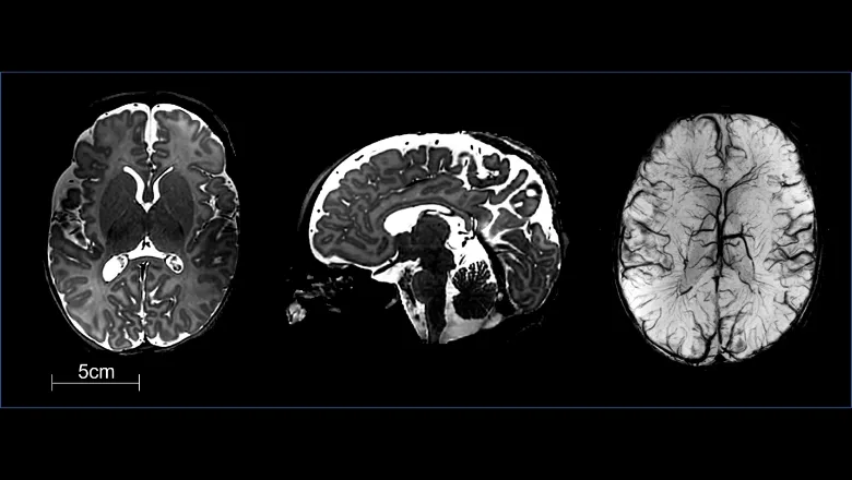
The researchers say they are excited about the extra detail seen on these early images in comparison to those acquired with a standard 1.5T or 3T MRI scanner. The quality of the scans will improve even more with further sequence optimisation. The images also highlight the possibilities for the technology moving forwards.
The increased signal-to-noise ratio the scanner offers means that clinicians and researchers can get high contrast, high resolution anatomical images such as these T2 images (which are at 0.4 microlitre resolution compared to typical images on standard scanners which are usually 1-2 microliters).
As a result, it will be possible to achieve much deeper characterisation of brain tissue development and pathology in a much more specific way than standard clinical field strengths.
This has implications not only for better disease identification, but also for understanding how a disease alters brain development.
The researchers say scanning at 7 Tesla will allow the use of particular MRI contrasts which greatly benefit from the stronger static magnetic field.
These include fMRI (which studies brain activity), Magnetic Resonance Spectroscopy (MRS) (which studies chemical levels in the brain), and susceptibility weighted imaging (SWI) (which can identify blood in blood vessels or from bleeding).
As these scanners are not built with neonatal imaging in mind, in order to get to this point the researchers had to prove that scanning babies would be safe. This involved a detailed safety study led by Dr Malik which was assisted by a Biomedical Engineering BEng student at King’s who helped to make a computer model of a neonate for their simulation work.
We are incredibly excited about acquiring these first images from babies on the 7T MRI scanner. Moving forwards, acquiring images from babies at 7T will present enormous opportunities for improving our ability to understand how the brain develops during the crucial first few weeks after birth and how this is altered in disease. It really advances clinical diagnosis and research in early human brain development.
Dr Tomoki Arichi, Clinical Reader in Perinatal Imaging
Claire Lemer, clinical director for medicine and neonatology at Evelina London Children’s Hospital, said: “Thanks to the research from our colleagues at King’s College London, we have started to use this new, advanced MRI scanner in our neonatology services. We’re so pleased to be able to use this new, advanced MRI scanner in clinical practice to help improve diagnosis and care that we offer to seriously ill and premature babies. Having maternity and neonatal care all under one roof, with the expertise of researchers at King’s College London, means that we can offer the very best and latest care for our women and babies. Having more detailed images of babies’ brains is so important for us in diagnosing and treating complex conditions, especially being able to see tiny features is crucial in looking after premature and tiny babies.”
In this story

Head of the Department of Early Life Imaging

Head of the Department of Imaging Physics and Engineering