The Nikon Imaging Centre (NIC) at King’s College London is a Core Facility for light microscopy, developed and operated as a partnership between King’s College London and Nikon Instruments UK.
This partnership allows us to offer the latest state-of-the-art light microscopy and imaging to the research community and the Centre serves as a learning platform for our regional corporate partners and contributors.
Our mission is to promote innovation across a broad range of biological research fields by providing access to cutting edge microscopy and imaging equipment.
Before requesting access to the Centre, please review the access policy and follow the steps outlined in the 'access' tab below to request an account for the online scheduling system and to arrange training.
We provide comprehensive training in basic and advanced light microscopy techniques and access to a broad range of Nikon microscopes from widefield to super-resolution. Nikon Imaging Centre staff support users on site and the Centre has close links to Advanced Imaging Specialists and Engineers at Nikon. We offer technical consultation and ongoing support to the research community at King’s College London and beyond to ensure researchers achieve high-quality data outputs.
We contribute to teaching and education at King’s College London as well as hosting instrument demos and workshops in partnership with Nikon for the benefit of the local imaging community.
Facility staff
Affiliated academics
Related equipment
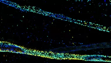
N-STORM super resolution
Super resolution - Imaging at lateral resolution of approximately 20nm, and axial of 50nm can be achieved.
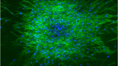
SoRa - spinning disk super-resolution by optical pixel reassignment
Fast confocal and super-resolution - equipped with a Yokogawa CSU-W1 dual disk scan head. High-speed, low phototoxicity confocal imaging possible.

AXR inverted confocal with NSPARC 1
Fast point scanning confocal-based imaging at depth. This microscope provides the option of super-resolution and gentle long-term imaging of live samples.

AXR inverted confocal with NSPARC 2
Fast point-scanning confocal imaging at depth. The NSPARC detector provides the option of super-resolution and gentle long-term imaging of live samples.

AXR inverted confocal
Fast point scanning confocal-based imaging at depth.

A1 upright confocal (with marzhauser multi slide stage for slide scanning)
High resolution imaging - The A1+ utilises a Galvano scanner, enabling high-resolution imaging of up to 4096 x 4096 pixels (512 x 512 pixels also possible).

AXR MP with NSPARC
Multiphoton confocal microscope, allowing for visualisation of fine structures deep within living organisms at high speeds

Widefield microscopes
Nikon's Ti2 inverted microscopes are fully motorised for multi-dimensional imaging, equipped with Nikon’s Perfect Focus System.

Eclipse JI
The Nikon Eclipse Ji is an all-digital smart imaging system.
Selected publications
2025
Picco, F. et al. (2025) ‘Macrophage to neuron communication via extracellular vesicles in neuropathic pain conditions’, Heliyon, 11(1). Available at: https://doi.org/10.1016/j.heliyon.2024.e41268.
2024
Aggarwal, N. et al. (2024) ‘Characterisation and correction of polarisation effects in fluorescently labelled fibres’, Journal of Microscopy [Preprint]. Available at: https://doi.org/10.1111/jmi.13308.
Cheung, A. et al. (2024) ‘Anti-EGFR Antibody–Drug Conjugate Carrying an Inhibitor Targeting CDK Restricts Triple-Negative Breast Cancer Growth’, Clinical Cancer Research, 30(15), pp. 3298–3315. Available at: https://doi.org/10.1158/1078-0432.CCR-23-3110.
Costanzo, H. et al. (2024) ‘The Development and Characterisation of ssDNA Aptamers via a Modified Cell-SELEX Methodology for the Detection of Human Red Blood Cells’, International Journal of Molecular Sciences, 25(3), p. 1814. Available at: https://doi.org/10.3390/ijms25031814.
Cronin, R. et al. (2024) ‘The p90 Ribosomal S6 Kinases 2 and 4 promote Prostate Cancer cell proliferation in androgen-dependent and independent ways’. bioRxiv, p. 2024.03.04.582739. Available at: https://doi.org/10.1101/2024.03.04.582739.
Culley, S. et al. (2024) ‘Made to measure: An introduction to quantifying microscopy data in the life sciences’, Journal of Microscopy, 295(1), pp. 61–82. Available at: https://doi.org/10.1111/jmi.13208.
Francis, L. et al. (2024) ‘Single-cell analysis of psoriasis resolution demonstrates an inflammatory fibroblast state targeted by IL-23 blockade’, Nature Communications, 15(1), p. 913. Available at: https://doi.org/10.1038/s41467-024-44994-w.
Konishi, K. et al. (2024) ‘Semi-automated navigation for efficient targeting of electron tomography to regions of interest in volume correlative light and electron microscopy’. bioRxiv, p. 2024.11.29.626074. Available at: https://doi.org/10.1101/2024.11.29.626074.
Lei, Y. et al. (2024) ‘Mapping the druggable targets displayed by human colonic enteroendocrine cells’. bioRxiv, p. 2024.10.29.620704. Available at: https://doi.org/10.1101/2024.10.29.620704.
Sofoluwe, A. et al. (2024) ‘Neutrophil NADPH oxidase breaks the inflammatory IL-1β/IL-17A circuit to enhance pathogen clearance during respiratory virus infections’. bioRxiv, p. 2024.09.12.612505. Available at: https://doi.org/10.1101/2024.09.12.612505.
2023
Alkattan, R. et al. (2023) ‘A self-etch bonding system with potential to eliminate selective etching and resist proteolytic degradation’, Journal of Dentistry, 132, p. 104501. Available at: https://doi.org/10.1016/j.jdent.2023.104501.
Anstee, J.E. et al. (2023) ‘LYVE-1+ macrophages form a collaborative CCR5-dependent perivascular niche that influences chemotherapy responses in murine breast cancer’, Developmental Cell, 58(17), pp. 1548-1561.e10. Available at: https://doi.org/10.1016/j.devcel.2023.06.006.
Carrington, G. et al. (2023) ‘Human skeletal myopathy myosin mutations disrupt myosin head sequestration’, JCI Insight, 8(21). Available at: https://doi.org/10.1172/jci.insight.172322.
Chandler, J. et al. (2023) ‘In situ FRET-based localization of the N terminus of myosin binding protein-C in heart muscle cells’, Proceedings of the National Academy of Sciences, 120(12), p. e2222005120. Available at: https://doi.org/10.1073/pnas.2222005120.
Crescioli, S. et al. (2023) ‘B cell profiles, antibody repertoire and reactivity reveal dysregulated responses with autoimmune features in melanoma’, Nature Communications, 14(1), p. 3378. Available at: https://doi.org/10.1038/s41467-023-39042-y.
Fanelli, G. et al. (2023) ‘Soluble Collectin 11 (CL-11) Acts as an Immunosuppressive Molecule Potentially Used by Stem Cell-Derived Retinal Epithelial Cells to Modulate T Cell Response’, Cells, 12(13), p. 1805. Available at: https://doi.org/10.3390/cells12131805.
Glover, J. et al. (2023) ‘UMAD1 contributes to ESCRT-III dynamic subunit turnover during cytokinetic abscission’, Journal of Cell Science, 136(15), p. jcs261097. Available at: https://doi.org/10.1242/jcs.261097.
Glebov, O.O. et al. (2023) ‘Structural synaptic signatures of Alzheimer’s disease and dementia with Lewy bodies in the male brain’, Neuropathology and Applied Neurobiology, 49(1), p. e12852. Available at: https://doi.org/10.1111/nan.12852.
Jahin, I. et al. (2023) ‘Extracellular matrix stiffness activates mechanosensitive signals but limits breast cancer cell spheroid proliferation and invasion’, Frontiers in Cell and Developmental Biology, 11. Available at: https://doi.org/10.3389/fcell.2023.1292775.
Patel, S. et al. (2023) ‘Programmed myofibre necrosis in critical illness acquired muscle wasting’, JCSM Rapid Communications, 6(2), pp. 111–121. Available at: https://doi.org/10.1002/rco2.83.
Perez-Ortiz, G. et al. (2023) ‘Production of copropophyrin III, biliverdin and bilirubin by the rufomycin producer, Streptomyces atratus’, Frontiers in Microbiology, 14. Available at: https://doi.org/10.3389/fmicb.2023.1092166.
Skourti, E. et al. (2023) ‘Spatiotemporal quantitative microRNA-155 imaging reports immune-mediated changes in a triple-negative breast cancer model’, Frontiers in Immunology, 14. Available at: https://doi.org/10.3389/fimmu.2023.1180233.
2022
Bourke, S. et al. (2022) ‘Biocompatible Magnetic Conjugated Polymer Nanoparticles for Optical and Lifetime Imaging Applications in the First Biological Window’, ACS Applied Polymer Materials, 4(11), pp. 8193–8202. Available at: https://doi.org/10.1021/acsapm.2c01153.
Donà, F. et al. (2022) ‘Removal of Stomatin, a Membrane-Associated Cell Division Protein, Results in Specific Cellular Lipid Changes’, Journal of the American Chemical Society, 144(39), pp. 18069–18074. Available at: https://doi.org/10.1021/jacs.2c07907.
Louis, B. et al. (2022) ‘A reductionist approach to determine the effect of cell-cell contact on human epidermal stem cell differentiation’, Acta Biomaterialia, 150, pp. 265–276. Available at: https://doi.org/10.1016/j.actbio.2022.07.054.
Parsons, R.B. et al. (2022) ‘Alpha-synucleinopathy reduces NMNAT3 protein levels and neurite formation that can be rescued by targeting the NAD+ pathway’, Human Molecular Genetics, 31(17), pp. 2918–2933. Available at: https://doi.org/10.1093/hmg/ddac077.
Ranu, N. et al. (2022) ‘NEB mutations disrupt the super-relaxed state of myosin and remodel the muscle metabolic proteome in nemaline myopathy’, Acta Neuropathologica Communications, 10(1), p. 185. Available at: https://doi.org/10.1186/s40478-022-01491-9.
2021
Fadul, J., Zulueta-Coarasa, T., Slattum, G.M. et al. (2021) KRas-transformed epithelia cells invade and partially dedifferentiate by basal cell extrusion. Nat Comm; 12, 7180.
Nikolaos Ioannou, Patrick R. Hagner, Matt Stokes, Anita K. Gandhi, Benedetta Apollonio, Mariam Fanous, Despoina Papazoglou, Lesley-Ann Sutton, Richard Rosenquist, Rose-Marie Amini, Hsiling Chiu, Antonia Lopez-Girona, Preethi Janardhanan, Farrukh T. Awan, Jeffrey Jones, Neil E. Kay, Tait D. Shanafelt, Martin S. Tallman, Kostas Stamatopoulos, Piers E. M. Patten, Anna Vardi, Alan G. Ramsay. (2021) Triggering interferon signaling in T cells with avadomide sensitizes CLL to anti-PD-L1/PD-1 immunotherapy.; 137 (2): 216–231.
Sioned F. Jones, Himanshu Joshi, Stephen J. Terry, Jonathan R. Burns, Aleksei Aksimentiev, Ulrike S. Eggert, and Stefan Howorka. (2021). Hydrophobic Interactions between DNA Duplexes and Synthetic and Biological Membranes. J. Am. Chem. Soc.; 143 (22), 8305-8313.
2020
Woods S, O'Brien LM, Butcher W, et al. (2020) Glucosamine-NISV delivers antibody across the blood-brain barrier: Optimization for treatment of encephalitic viruses. J Control Release; 324: 644-656.
Michael J. Shannon, Judith Pineau, Juliette Griffié, Jesse Aaron, Tamlyn Peel, David J. Williamson, Rose Zamoyska, Andrew P. Cope, Georgina H. Cornish, Dylan M. Owen, Ana-Maria Lennon-Duménil. (2020). Differential nanoscale organisation of LFA-1 modulates T-cell migration. J Cell Sci; 133 (5): jcs232991.
2019
Negri VA, Logtenberg MEW, Renz LM, Oules B, Walko G, Watt FM. (2019). Delta-like 1-mediated cis-inhibition of Jagged1/2 signalling inhibits differentiation of human epidermal cells in culture. Sci Rep. 9 (1):10825.
Morton PE, Perrin C, Levitt J, et al. (2019). TNFR1 membrane reorganization promotes distinct modes of TNFα signaling. Sci Signal. 12(592):eaaw2418.
Ross JA, Levy Y, Ripolone M, et al (2019). Impairments in contractility and cytoskeletal organisation cause nuclear defects in nemaline myopathy. Acta Neuropathol;138(3):477-495.
Evans R, Flores-Borja F, Nassiri S, et al. (2019). Integrin-Mediated Macrophage Adhesion Promotes Lymphovascular Dissemination in Breast Cancer. Cell Rep; 27(7):1967-1978.e4.
Ventimiglia LN, Cuesta-Geijo MA, Martinelli N, et al (2018). CC2D1B Coordinates ESCRT-III Activity during the Mitotic Reformation of the Nuclear Envelope. Dev Cell. 47 (5):547-563.e6.
Doyle T, Moncorgé O, Bonaventure B, et al. (2018). The interferon-inducible isoform of NCOA7 inhibits endosome-mediated viral entry. Nat Microbiol., 4(3):539].
2018
Sadler JBA, Wenzel DM, Williams LK, et al. (2018). A cancer-associated polymorphism in ESCRT-III disrupts the abscission checkpoint and promotes genome instability. Proc Natl Acad Sci U S A. 115(38):E8900-E8908.
Michael M, Begum R, Chan GK, et al. (2019). Kindlin-1 Regulates Epidermal Growth Factor Receptor Signaling. J Invest Dermatol;139(2):369-379.
Rognoni E, Pisco AO, Hiratsuka T, et al. (2018). Fibroblast state switching orchestrates dermal maturation and wound healing. Mol Syst Biol.14(8):e8174.
Cheung A, Opzoomer J, Ilieva KM, et al. (2018). Anti-Folate Receptor Alpha-Directed Antibody Therapies Restrict the Growth of Triple-negative Breast Cancer. Clin Cancer Res. 24(20):5098-5111.
Monypenny J, Milewicz H, Flores-Borja F, et al. (2018). ALIX Regulates Tumor-Mediated Immunosuppression by Controlling EGFR Activity and PD-L1 Presentation. Cell Rep. 24(3):630-641.
Marsh R.J, Pfisterer K, Bennett P, Hirvonen LM, Gautel M, Jones GE, Cox S. (2018). Artifact-free high-density localization microscopy analysis. Nature Methods.
Peters R, Griffié J, Burn GL, Williamson DJ, Owen DM. (2018). Quantitative fibre analysis of single-molecule localization microscopy data. Scientific Reports. 8; 10418.
2017
Ashdown GW, Burn GL, Williamson DJ, Pandžić E, Peters R, Holden M, Ewers H, Shao L, Wiseman PW, Owen DM. (2017). Live-Cell Super-resolution Reveals F-Actin and Plasma Membrane Dynamics at the T Cell Synapse. Biophysical Journal. 112;1703–1713.
2016
Starling GP, Yip YY, Sanger A, Morton PE, Eden ER, Dodding MP. (2016). Folliculin directs the formation of a Rab34–RILP complex to control the nutrient‐dependent dynamic distribution of lysosomes. EMBO Reports. 17; 823-841
Deans PJM, Raval P, Sellers KJ, Gatford NJF, Halai S, Duarte RRR, Shum C, Warre-Cornish K, Kaplun VE, Cocks G, Hill M, Bray NJ, Price J, Srivastava DP. (2016). Psychosis risk candidate ZNF804A localizes to synapses and regulates neurite formation and dendritic spine structure. Biological Psychiatry
Olmos Y, Perdrix-Rosell A, Carlton JG. (2016). Membrane Binding by CHMP7 Coordinates ESCRT-III-Dependent Nuclear Envelope Reformation. Current Biology.
Jayo A, Malboubi M, Antoku S, Chang W, Ortiz-Zapater E, Groen C, Pfisterer K, Tootle T, Charras G, Gundersen GG, Parsons M. (2016) Regulates Nuclear Movement and Deformation in Migrating Cells. Developmental Cell. 38(4):371-383.
Arbore G, West EE, Spolski R, Robertson AAB, Klos A, Rheinheimer C, Dutow P, Woodruff TM, Yu ZX, O’Neill LA, Coll RC, Sher A, Leonard WJ, Köhl J, Monk P, Cooper MA, Arno M, Afzali B, Lachmann HJ, Cope AP, Mayer-Barber KD, Kemper C. (2016). T helper 1 immunity requires complement-driven NLRP3 inflammasome activity in CD4+ T cells. Science. 352; 6292.
2015
Villari G, Jayo A, Zanet J, Fitch B, Serrels B, Frame M, Stramer B, Goult BT, Parsons M. (2015). A direct interaction between fascin and microtubules contributes to adhesion dynamics and cell migration. J Cell Sci. 128(24):4601-14
2014
Ashdown GW, Cope A, Wiseman PW, Owen DM. (2014). Molecular Flow Quantified beyond the Diffraction Limit by Spatiotemporal Image Correlation of Structured Illumination Microscopy Data. Biophysical Journal, 107(9), L21–L23.
Marra P, Mathew S, Grigoriadis A, Wu Y, Kyle-Cezar F, Watkins J, Rashid M, De Rinaldis E, Hessey S, Gazinska P, Hayday A, Tutt A. (2014). IL15RA Drives Antagonistic Mechanisms of Cancer Development and Immune Control in Lymphocyte-Enriched Triple-Negative Breast Cancers. Cancer Research.74(17)
2013
Stürzenbaum SR, Höckner M, Panneerselvam A, Levitt J, Bouillard JS, Taniguchi S, Dailey LA, Ahmad Khanbeigi R, Rosca EV, Thanou M et al. (2013). Biosynthesis of luminescent quantum dots in an earthworm. Nature Nanotechnology 8, 57–60.
Hng KI, Dormann D. (2013). ConfocalCheck - A Software Tool for the Automated Monitoring of Confocal Microscope Performance. PLoS ONE 8, e79879.
Asgari, E., Le Friec, G., Yamamoto, H., Perucha, E., Sacks, S.S., Köhl, J., Cook, H.T., and Kemper, C. (2013). C3a modulates IL-1β secretion in human monocytes by regulating ATP efflux and subsequent NLRP3 inflammasome activation. Blood. 122(20):3473-81
The Centre is committed to collaborative research and innovation through open access to our state-of-the-art core facilities. We welcome King’s researchers, external academic collaborators commercial clients and industry partners.
Access rates
- Rates vary depending on funding source. Please contact the team for our 2025 access rates.
King’s College London researchers
- Current facility users can book instruments and services via King’s our online booking system.
- Prospective users must submit two forms
Account Request Form - Include a valid grant code in the ‘Account Number’ field
Training Request Form - Submit one form per microscope, specifying sample details and instrument preferences. Please refer to the NIC access policy.
- Training - Two sessions are required before independent use:
Session 1: Demonstration and setup (bring samples if possible).
Session 2: Hands-on operation under supervision.
External academic, commercial clients and industry partners
The Nikon Imaging Centre (NIC) welcomes external academic researchers, commercial clients, and industry partners.
We offer flexible access models and tailored collaboration opportunities to support a wide range of research and innovation needs. Whether you're seeking imaging support, joint research initiatives, or long-term partnerships, NIC is equipped to help.
For access enquiries, service details, collaboration opportunities, or quotations, please contact the NIC team.
NIC opening times
- Weekdays, 09:00 –17:00
- Out-of-hours access is available upon request once competency is demonstrated.
We provide comprehensive training in basic and advanced light microscopy techniques and access to a broad range of Nikon microscopes from widefield to super-resolution. Nikon Imaging Centre staff support users on site and the Centre has close links to Advanced Imaging Specialists and Engineers at Nikon. We offer technical consultation and ongoing support to the research community at King’s College London and beyond to ensure researchers achieve high-quality data outputs.
We contribute to teaching and education at King’s College London as well as hosting instrument demos and workshops in partnership with Nikon for the benefit of the local imaging community.
Facility staff
Affiliated academics
Related equipment

N-STORM super resolution
Super resolution - Imaging at lateral resolution of approximately 20nm, and axial of 50nm can be achieved.

SoRa - spinning disk super-resolution by optical pixel reassignment
Fast confocal and super-resolution - equipped with a Yokogawa CSU-W1 dual disk scan head. High-speed, low phototoxicity confocal imaging possible.

AXR inverted confocal with NSPARC 1
Fast point scanning confocal-based imaging at depth. This microscope provides the option of super-resolution and gentle long-term imaging of live samples.

AXR inverted confocal with NSPARC 2
Fast point-scanning confocal imaging at depth. The NSPARC detector provides the option of super-resolution and gentle long-term imaging of live samples.

AXR inverted confocal
Fast point scanning confocal-based imaging at depth.

A1 upright confocal (with marzhauser multi slide stage for slide scanning)
High resolution imaging - The A1+ utilises a Galvano scanner, enabling high-resolution imaging of up to 4096 x 4096 pixels (512 x 512 pixels also possible).

AXR MP with NSPARC
Multiphoton confocal microscope, allowing for visualisation of fine structures deep within living organisms at high speeds

Widefield microscopes
Nikon's Ti2 inverted microscopes are fully motorised for multi-dimensional imaging, equipped with Nikon’s Perfect Focus System.

Eclipse JI
The Nikon Eclipse Ji is an all-digital smart imaging system.
Selected publications
2025
Picco, F. et al. (2025) ‘Macrophage to neuron communication via extracellular vesicles in neuropathic pain conditions’, Heliyon, 11(1). Available at: https://doi.org/10.1016/j.heliyon.2024.e41268.
2024
Aggarwal, N. et al. (2024) ‘Characterisation and correction of polarisation effects in fluorescently labelled fibres’, Journal of Microscopy [Preprint]. Available at: https://doi.org/10.1111/jmi.13308.
Cheung, A. et al. (2024) ‘Anti-EGFR Antibody–Drug Conjugate Carrying an Inhibitor Targeting CDK Restricts Triple-Negative Breast Cancer Growth’, Clinical Cancer Research, 30(15), pp. 3298–3315. Available at: https://doi.org/10.1158/1078-0432.CCR-23-3110.
Costanzo, H. et al. (2024) ‘The Development and Characterisation of ssDNA Aptamers via a Modified Cell-SELEX Methodology for the Detection of Human Red Blood Cells’, International Journal of Molecular Sciences, 25(3), p. 1814. Available at: https://doi.org/10.3390/ijms25031814.
Cronin, R. et al. (2024) ‘The p90 Ribosomal S6 Kinases 2 and 4 promote Prostate Cancer cell proliferation in androgen-dependent and independent ways’. bioRxiv, p. 2024.03.04.582739. Available at: https://doi.org/10.1101/2024.03.04.582739.
Culley, S. et al. (2024) ‘Made to measure: An introduction to quantifying microscopy data in the life sciences’, Journal of Microscopy, 295(1), pp. 61–82. Available at: https://doi.org/10.1111/jmi.13208.
Francis, L. et al. (2024) ‘Single-cell analysis of psoriasis resolution demonstrates an inflammatory fibroblast state targeted by IL-23 blockade’, Nature Communications, 15(1), p. 913. Available at: https://doi.org/10.1038/s41467-024-44994-w.
Konishi, K. et al. (2024) ‘Semi-automated navigation for efficient targeting of electron tomography to regions of interest in volume correlative light and electron microscopy’. bioRxiv, p. 2024.11.29.626074. Available at: https://doi.org/10.1101/2024.11.29.626074.
Lei, Y. et al. (2024) ‘Mapping the druggable targets displayed by human colonic enteroendocrine cells’. bioRxiv, p. 2024.10.29.620704. Available at: https://doi.org/10.1101/2024.10.29.620704.
Sofoluwe, A. et al. (2024) ‘Neutrophil NADPH oxidase breaks the inflammatory IL-1β/IL-17A circuit to enhance pathogen clearance during respiratory virus infections’. bioRxiv, p. 2024.09.12.612505. Available at: https://doi.org/10.1101/2024.09.12.612505.
2023
Alkattan, R. et al. (2023) ‘A self-etch bonding system with potential to eliminate selective etching and resist proteolytic degradation’, Journal of Dentistry, 132, p. 104501. Available at: https://doi.org/10.1016/j.jdent.2023.104501.
Anstee, J.E. et al. (2023) ‘LYVE-1+ macrophages form a collaborative CCR5-dependent perivascular niche that influences chemotherapy responses in murine breast cancer’, Developmental Cell, 58(17), pp. 1548-1561.e10. Available at: https://doi.org/10.1016/j.devcel.2023.06.006.
Carrington, G. et al. (2023) ‘Human skeletal myopathy myosin mutations disrupt myosin head sequestration’, JCI Insight, 8(21). Available at: https://doi.org/10.1172/jci.insight.172322.
Chandler, J. et al. (2023) ‘In situ FRET-based localization of the N terminus of myosin binding protein-C in heart muscle cells’, Proceedings of the National Academy of Sciences, 120(12), p. e2222005120. Available at: https://doi.org/10.1073/pnas.2222005120.
Crescioli, S. et al. (2023) ‘B cell profiles, antibody repertoire and reactivity reveal dysregulated responses with autoimmune features in melanoma’, Nature Communications, 14(1), p. 3378. Available at: https://doi.org/10.1038/s41467-023-39042-y.
Fanelli, G. et al. (2023) ‘Soluble Collectin 11 (CL-11) Acts as an Immunosuppressive Molecule Potentially Used by Stem Cell-Derived Retinal Epithelial Cells to Modulate T Cell Response’, Cells, 12(13), p. 1805. Available at: https://doi.org/10.3390/cells12131805.
Glover, J. et al. (2023) ‘UMAD1 contributes to ESCRT-III dynamic subunit turnover during cytokinetic abscission’, Journal of Cell Science, 136(15), p. jcs261097. Available at: https://doi.org/10.1242/jcs.261097.
Glebov, O.O. et al. (2023) ‘Structural synaptic signatures of Alzheimer’s disease and dementia with Lewy bodies in the male brain’, Neuropathology and Applied Neurobiology, 49(1), p. e12852. Available at: https://doi.org/10.1111/nan.12852.
Jahin, I. et al. (2023) ‘Extracellular matrix stiffness activates mechanosensitive signals but limits breast cancer cell spheroid proliferation and invasion’, Frontiers in Cell and Developmental Biology, 11. Available at: https://doi.org/10.3389/fcell.2023.1292775.
Patel, S. et al. (2023) ‘Programmed myofibre necrosis in critical illness acquired muscle wasting’, JCSM Rapid Communications, 6(2), pp. 111–121. Available at: https://doi.org/10.1002/rco2.83.
Perez-Ortiz, G. et al. (2023) ‘Production of copropophyrin III, biliverdin and bilirubin by the rufomycin producer, Streptomyces atratus’, Frontiers in Microbiology, 14. Available at: https://doi.org/10.3389/fmicb.2023.1092166.
Skourti, E. et al. (2023) ‘Spatiotemporal quantitative microRNA-155 imaging reports immune-mediated changes in a triple-negative breast cancer model’, Frontiers in Immunology, 14. Available at: https://doi.org/10.3389/fimmu.2023.1180233.
2022
Bourke, S. et al. (2022) ‘Biocompatible Magnetic Conjugated Polymer Nanoparticles for Optical and Lifetime Imaging Applications in the First Biological Window’, ACS Applied Polymer Materials, 4(11), pp. 8193–8202. Available at: https://doi.org/10.1021/acsapm.2c01153.
Donà, F. et al. (2022) ‘Removal of Stomatin, a Membrane-Associated Cell Division Protein, Results in Specific Cellular Lipid Changes’, Journal of the American Chemical Society, 144(39), pp. 18069–18074. Available at: https://doi.org/10.1021/jacs.2c07907.
Louis, B. et al. (2022) ‘A reductionist approach to determine the effect of cell-cell contact on human epidermal stem cell differentiation’, Acta Biomaterialia, 150, pp. 265–276. Available at: https://doi.org/10.1016/j.actbio.2022.07.054.
Parsons, R.B. et al. (2022) ‘Alpha-synucleinopathy reduces NMNAT3 protein levels and neurite formation that can be rescued by targeting the NAD+ pathway’, Human Molecular Genetics, 31(17), pp. 2918–2933. Available at: https://doi.org/10.1093/hmg/ddac077.
Ranu, N. et al. (2022) ‘NEB mutations disrupt the super-relaxed state of myosin and remodel the muscle metabolic proteome in nemaline myopathy’, Acta Neuropathologica Communications, 10(1), p. 185. Available at: https://doi.org/10.1186/s40478-022-01491-9.
2021
Fadul, J., Zulueta-Coarasa, T., Slattum, G.M. et al. (2021) KRas-transformed epithelia cells invade and partially dedifferentiate by basal cell extrusion. Nat Comm; 12, 7180.
Nikolaos Ioannou, Patrick R. Hagner, Matt Stokes, Anita K. Gandhi, Benedetta Apollonio, Mariam Fanous, Despoina Papazoglou, Lesley-Ann Sutton, Richard Rosenquist, Rose-Marie Amini, Hsiling Chiu, Antonia Lopez-Girona, Preethi Janardhanan, Farrukh T. Awan, Jeffrey Jones, Neil E. Kay, Tait D. Shanafelt, Martin S. Tallman, Kostas Stamatopoulos, Piers E. M. Patten, Anna Vardi, Alan G. Ramsay. (2021) Triggering interferon signaling in T cells with avadomide sensitizes CLL to anti-PD-L1/PD-1 immunotherapy.; 137 (2): 216–231.
Sioned F. Jones, Himanshu Joshi, Stephen J. Terry, Jonathan R. Burns, Aleksei Aksimentiev, Ulrike S. Eggert, and Stefan Howorka. (2021). Hydrophobic Interactions between DNA Duplexes and Synthetic and Biological Membranes. J. Am. Chem. Soc.; 143 (22), 8305-8313.
2020
Woods S, O'Brien LM, Butcher W, et al. (2020) Glucosamine-NISV delivers antibody across the blood-brain barrier: Optimization for treatment of encephalitic viruses. J Control Release; 324: 644-656.
Michael J. Shannon, Judith Pineau, Juliette Griffié, Jesse Aaron, Tamlyn Peel, David J. Williamson, Rose Zamoyska, Andrew P. Cope, Georgina H. Cornish, Dylan M. Owen, Ana-Maria Lennon-Duménil. (2020). Differential nanoscale organisation of LFA-1 modulates T-cell migration. J Cell Sci; 133 (5): jcs232991.
2019
Negri VA, Logtenberg MEW, Renz LM, Oules B, Walko G, Watt FM. (2019). Delta-like 1-mediated cis-inhibition of Jagged1/2 signalling inhibits differentiation of human epidermal cells in culture. Sci Rep. 9 (1):10825.
Morton PE, Perrin C, Levitt J, et al. (2019). TNFR1 membrane reorganization promotes distinct modes of TNFα signaling. Sci Signal. 12(592):eaaw2418.
Ross JA, Levy Y, Ripolone M, et al (2019). Impairments in contractility and cytoskeletal organisation cause nuclear defects in nemaline myopathy. Acta Neuropathol;138(3):477-495.
Evans R, Flores-Borja F, Nassiri S, et al. (2019). Integrin-Mediated Macrophage Adhesion Promotes Lymphovascular Dissemination in Breast Cancer. Cell Rep; 27(7):1967-1978.e4.
Ventimiglia LN, Cuesta-Geijo MA, Martinelli N, et al (2018). CC2D1B Coordinates ESCRT-III Activity during the Mitotic Reformation of the Nuclear Envelope. Dev Cell. 47 (5):547-563.e6.
Doyle T, Moncorgé O, Bonaventure B, et al. (2018). The interferon-inducible isoform of NCOA7 inhibits endosome-mediated viral entry. Nat Microbiol., 4(3):539].
2018
Sadler JBA, Wenzel DM, Williams LK, et al. (2018). A cancer-associated polymorphism in ESCRT-III disrupts the abscission checkpoint and promotes genome instability. Proc Natl Acad Sci U S A. 115(38):E8900-E8908.
Michael M, Begum R, Chan GK, et al. (2019). Kindlin-1 Regulates Epidermal Growth Factor Receptor Signaling. J Invest Dermatol;139(2):369-379.
Rognoni E, Pisco AO, Hiratsuka T, et al. (2018). Fibroblast state switching orchestrates dermal maturation and wound healing. Mol Syst Biol.14(8):e8174.
Cheung A, Opzoomer J, Ilieva KM, et al. (2018). Anti-Folate Receptor Alpha-Directed Antibody Therapies Restrict the Growth of Triple-negative Breast Cancer. Clin Cancer Res. 24(20):5098-5111.
Monypenny J, Milewicz H, Flores-Borja F, et al. (2018). ALIX Regulates Tumor-Mediated Immunosuppression by Controlling EGFR Activity and PD-L1 Presentation. Cell Rep. 24(3):630-641.
Marsh R.J, Pfisterer K, Bennett P, Hirvonen LM, Gautel M, Jones GE, Cox S. (2018). Artifact-free high-density localization microscopy analysis. Nature Methods.
Peters R, Griffié J, Burn GL, Williamson DJ, Owen DM. (2018). Quantitative fibre analysis of single-molecule localization microscopy data. Scientific Reports. 8; 10418.
2017
Ashdown GW, Burn GL, Williamson DJ, Pandžić E, Peters R, Holden M, Ewers H, Shao L, Wiseman PW, Owen DM. (2017). Live-Cell Super-resolution Reveals F-Actin and Plasma Membrane Dynamics at the T Cell Synapse. Biophysical Journal. 112;1703–1713.
2016
Starling GP, Yip YY, Sanger A, Morton PE, Eden ER, Dodding MP. (2016). Folliculin directs the formation of a Rab34–RILP complex to control the nutrient‐dependent dynamic distribution of lysosomes. EMBO Reports. 17; 823-841
Deans PJM, Raval P, Sellers KJ, Gatford NJF, Halai S, Duarte RRR, Shum C, Warre-Cornish K, Kaplun VE, Cocks G, Hill M, Bray NJ, Price J, Srivastava DP. (2016). Psychosis risk candidate ZNF804A localizes to synapses and regulates neurite formation and dendritic spine structure. Biological Psychiatry
Olmos Y, Perdrix-Rosell A, Carlton JG. (2016). Membrane Binding by CHMP7 Coordinates ESCRT-III-Dependent Nuclear Envelope Reformation. Current Biology.
Jayo A, Malboubi M, Antoku S, Chang W, Ortiz-Zapater E, Groen C, Pfisterer K, Tootle T, Charras G, Gundersen GG, Parsons M. (2016) Regulates Nuclear Movement and Deformation in Migrating Cells. Developmental Cell. 38(4):371-383.
Arbore G, West EE, Spolski R, Robertson AAB, Klos A, Rheinheimer C, Dutow P, Woodruff TM, Yu ZX, O’Neill LA, Coll RC, Sher A, Leonard WJ, Köhl J, Monk P, Cooper MA, Arno M, Afzali B, Lachmann HJ, Cope AP, Mayer-Barber KD, Kemper C. (2016). T helper 1 immunity requires complement-driven NLRP3 inflammasome activity in CD4+ T cells. Science. 352; 6292.
2015
Villari G, Jayo A, Zanet J, Fitch B, Serrels B, Frame M, Stramer B, Goult BT, Parsons M. (2015). A direct interaction between fascin and microtubules contributes to adhesion dynamics and cell migration. J Cell Sci. 128(24):4601-14
2014
Ashdown GW, Cope A, Wiseman PW, Owen DM. (2014). Molecular Flow Quantified beyond the Diffraction Limit by Spatiotemporal Image Correlation of Structured Illumination Microscopy Data. Biophysical Journal, 107(9), L21–L23.
Marra P, Mathew S, Grigoriadis A, Wu Y, Kyle-Cezar F, Watkins J, Rashid M, De Rinaldis E, Hessey S, Gazinska P, Hayday A, Tutt A. (2014). IL15RA Drives Antagonistic Mechanisms of Cancer Development and Immune Control in Lymphocyte-Enriched Triple-Negative Breast Cancers. Cancer Research.74(17)
2013
Stürzenbaum SR, Höckner M, Panneerselvam A, Levitt J, Bouillard JS, Taniguchi S, Dailey LA, Ahmad Khanbeigi R, Rosca EV, Thanou M et al. (2013). Biosynthesis of luminescent quantum dots in an earthworm. Nature Nanotechnology 8, 57–60.
Hng KI, Dormann D. (2013). ConfocalCheck - A Software Tool for the Automated Monitoring of Confocal Microscope Performance. PLoS ONE 8, e79879.
Asgari, E., Le Friec, G., Yamamoto, H., Perucha, E., Sacks, S.S., Köhl, J., Cook, H.T., and Kemper, C. (2013). C3a modulates IL-1β secretion in human monocytes by regulating ATP efflux and subsequent NLRP3 inflammasome activation. Blood. 122(20):3473-81
The Centre is committed to collaborative research and innovation through open access to our state-of-the-art core facilities. We welcome King’s researchers, external academic collaborators commercial clients and industry partners.
Access rates
- Rates vary depending on funding source. Please contact the team for our 2025 access rates.
King’s College London researchers
- Current facility users can book instruments and services via King’s our online booking system.
- Prospective users must submit two forms
Account Request Form - Include a valid grant code in the ‘Account Number’ field
Training Request Form - Submit one form per microscope, specifying sample details and instrument preferences. Please refer to the NIC access policy.
- Training - Two sessions are required before independent use:
Session 1: Demonstration and setup (bring samples if possible).
Session 2: Hands-on operation under supervision.
External academic, commercial clients and industry partners
The Nikon Imaging Centre (NIC) welcomes external academic researchers, commercial clients, and industry partners.
We offer flexible access models and tailored collaboration opportunities to support a wide range of research and innovation needs. Whether you're seeking imaging support, joint research initiatives, or long-term partnerships, NIC is equipped to help.
For access enquiries, service details, collaboration opportunities, or quotations, please contact the NIC team.
NIC opening times
- Weekdays, 09:00 –17:00
- Out-of-hours access is available upon request once competency is demonstrated.
Partners

Contact us
3rd Floor, Hodgkin Building Guys Campus King’s College London London, SE1 1UL

