Welcome to The Spatial Biology Facility |
|
A joint initiative between the Faculty of Life Sciences & Medicine and the Faculty of Dentistry, Oral & Craniofacial Sciences at King’s College London
Our Facility
We provide our research community with spatial transcriptomics, proteomics and multi-omics solutions to assist with their research projects.
Our Goal
With a strong focus on producing accurate, reproducible data and the bioinformatics support to interpret the results, we aim to foster a collaborative environment across disciplines so that investigators can innovate and transform their research.

Single-Cell Spatial Omics


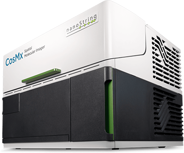
NanoString CosMx SMI
The CosMx Spatial Molecular Imager (SMI) enables high-plex spatial profiling, allowing quantification and visualization of 1,000, 6,175, or the whole transcriptome (~19,000 transcripts). For proteomic analysis, it supports 64 validated human proteins and 68 mouse proteins (for neuroscience applications), with the option to include up to 8 customizable protein targets.
The platform also enables multiomics by combining protein and RNA (at any of the three plex levels) analysis on the same tissue section, allowing researchers to comprehensively map single cells within their spatial context.
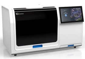
10x Genomics Xenium Analyzer
The 10x Genomics Xenium Analyzer enables bespoke spatial analysis using custom-made panels (480 transcripts), pre-designed tissue-specific panels (up to 380 genes) or with the Human or Mouse Pan Tissue & Pathways Panel (5,006 transcripts). Additionally, up to 100 extra genes can be added to further customize the assay.
The multiomic capability of the platform is achieved by combining RNA spatial profiling (<500-gene panels) with protein detection (up to 27 targets) on the same tissue section.
Bioinformatics Analysis
Our extensive experience in spatial transcriptomics and proteomics enables us to support our customers.
- Tailored experimental designs
- In-depth bioinformatics analysis
- Data interpretation
Facility staff

Professor of Molecular and Digital Pathology

Reader in BioNano Engineering

Postdoctoral Research Fellow

Spatial Biology Facility Manager
- Alberts, E., et al. Extrafollicular plasma cells disable dendritic cell-T-cell priming in tumor-draining lymph nodes. Preprint (2026).
- Baptista, A., Cruiziat, D., Nuamah, R., Chiappini, C. & Grigoriadis, A. MOSAIK: Multi-Origin Spatial Transcriptomics Analysis and Integration Kit. J. Open Source Softw. 11, 8795 (2026).
- Chen, S., et al. Normal breast tissue (NBT)-classifiers: advancing compartment classification in normal breast histology. Npj Breast Cancer 12, 41 (2026).
- Durham, L. E., et al. Linking Skin and Joint Inflammation in Psoriatic Arthritis through Shared CD8+ T Cell Clones. Arthritis Rheumatol. 78, 152–165 (2026).
- Wall, I., Baptista, A., et al. SMART: A Spatio-Molecular Atlas of Response Trajectories in Triple-Negative Breast Cancer. Preprint (2025).
- Goss, G., et al. Mesodermal-niche interactions direct specification and differentiation of pancreatic islet cells in human multilineage organoids. Preprint (2025).
- Sasiain, I., et al. Genome-Wide DNA Methylation Profiling of Triple-Negative Breast Cancer Uncovers Epigenetic Biomarkers of Tumor Identity and the Immune Microenvironment. Preprint (2025).
- Gu, C., et al. Nanoneedles enable spatiotemporal lipidomics of living tissues. Nat. Nanotechnol. 20, 1262–1272 (2025).
- O’Hora, B., et al. NoButter: An R package for reducing transcript dispersion in CosMx Spatial Molecular Imaging Data. Preprint (2024).
- Lau, H. S. H., et al. Adipose-enriched peri-tumoral stroma, in contrast to myofibroblast-enriched stroma, prognosticates poorer survival in breast cancers. Npj Breast Cancer 9, 84 (2023).
- Li, M., et al. B Cells in Breast Cancer Pathology. Cancers 15, 1517 (2023).
- Verghese, G., et al. Computational pathology in cancer diagnosis, prognosis, and prediction – present day and prospects. J. Pathol. 260, 551–563 (2023).
- Verghese, G., et al. Multiscale deep learning framework captures systemic immune features in lymph nodes predictive of triple negative breast cancer outcome in large-scale studies. J. Pathol. 260, 376–389 (2023).
KHP Cancer Biobank
The King’s Health Partners Cancer Biobank is fundamental to our aim, as a Comprehensive Cancer Centre, of bringing together our worldclass research, teaching and clinical practice for the benefit of patients. We have a well-established, tradition of biobanking with a long history of tissue and data use across the UK and Europe. We are constantly improving our resources by collecting a more diverse range of sample types from the majority of Cancers. The Biobank resources are essential in understanding what causes Cancer, which treatment options are likely to be effective and how to improve patient care in the years to come.
The Hub for Applied Bioinformatics (HAB)
Welcome to the Hub for Applied Bioinformatics at KCL, a place where big data are transformed into valuable knowledge that can drive scientific breakthroughs in the fields of life sciences and medicine. Our mission is to create a collaborative environment where ideas and expertise can be shared freely, leading to greater productivity and innovation in the field of bioinformatics. Whether you're a student, a research associate or a PI, HAB is the perfect place to learn more about bioinformatics and its application and to getting research done. At the HAB you'll be able to apply for bioinformatics support, attend seminars and data club designed around research work streams, and attend training sessions taught by experts in the fields.

London Metallomics Facility
The London Metallomics Facility (LMF) is a King’s College London core research facility, providing state-of-the-art analytical tools to understand the critical roles of metals in biological processes. Our vision is to be a centralised hub for comprehensive multi-institutional integration of metallomic analytics, establishing direct interactions between leading research groups, commercial partners, and industry, whilst driving educational outreach and public engagement strategies.
NanoString CosMx SMI
The CosMx Spatial Molecular Imager (SMI) enables high-plex spatial profiling, allowing quantification and visualization of 1,000, 6,175, or the whole transcriptome (~19,000 transcripts). For proteomic analysis, it supports 64 validated human proteins and 68 mouse proteins (for neuroscience applications), with the option to include up to 8 customizable protein targets.
The platform also enables multiomics by combining protein and RNA (at any of the three plex levels) analysis on the same tissue section, allowing researchers to comprehensively map single cells within their spatial context.
10x Genomics Xenium Analyzer
The 10x Genomics Xenium Analyzer enables bespoke spatial analysis using custom-made panels (480 transcripts), pre-designed tissue-specific panels (up to 380 genes) or with the Human or Mouse Pan Tissue & Pathways Panel (5,006 transcripts). Additionally, up to 100 extra genes can be added to further customize the assay.
The multiomic capability of the platform is achieved by combining RNA spatial profiling (<500-gene panels) with protein detection (up to 27 targets) on the same tissue section.
Bioinformatics Analysis
Our extensive experience in spatial transcriptomics and proteomics enables us to support our customers.
- Tailored experimental designs
- In-depth bioinformatics analysis
- Data interpretation
Facility staff
Professor of Molecular and Digital Pathology
Reader in BioNano Engineering
Postdoctoral Research Fellow
Spatial Biology Facility Manager
- Alberts, E., et al. Extrafollicular plasma cells disable dendritic cell-T-cell priming in tumor-draining lymph nodes. Preprint (2026).
- Baptista, A., Cruiziat, D., Nuamah, R., Chiappini, C. & Grigoriadis, A. MOSAIK: Multi-Origin Spatial Transcriptomics Analysis and Integration Kit. J. Open Source Softw. 11, 8795 (2026).
- Chen, S., et al. Normal breast tissue (NBT)-classifiers: advancing compartment classification in normal breast histology. Npj Breast Cancer 12, 41 (2026).
- Durham, L. E., et al. Linking Skin and Joint Inflammation in Psoriatic Arthritis through Shared CD8+ T Cell Clones. Arthritis Rheumatol. 78, 152–165 (2026).
- Wall, I., Baptista, A., et al. SMART: A Spatio-Molecular Atlas of Response Trajectories in Triple-Negative Breast Cancer. Preprint (2025).
- Goss, G., et al. Mesodermal-niche interactions direct specification and differentiation of pancreatic islet cells in human multilineage organoids. Preprint (2025).
- Sasiain, I., et al. Genome-Wide DNA Methylation Profiling of Triple-Negative Breast Cancer Uncovers Epigenetic Biomarkers of Tumor Identity and the Immune Microenvironment. Preprint (2025).
- Gu, C., et al. Nanoneedles enable spatiotemporal lipidomics of living tissues. Nat. Nanotechnol. 20, 1262–1272 (2025).
- O’Hora, B., et al. NoButter: An R package for reducing transcript dispersion in CosMx Spatial Molecular Imaging Data. Preprint (2024).
- Lau, H. S. H., et al. Adipose-enriched peri-tumoral stroma, in contrast to myofibroblast-enriched stroma, prognosticates poorer survival in breast cancers. Npj Breast Cancer 9, 84 (2023).
- Li, M., et al. B Cells in Breast Cancer Pathology. Cancers 15, 1517 (2023).
- Verghese, G., et al. Computational pathology in cancer diagnosis, prognosis, and prediction – present day and prospects. J. Pathol. 260, 551–563 (2023).
- Verghese, G., et al. Multiscale deep learning framework captures systemic immune features in lymph nodes predictive of triple negative breast cancer outcome in large-scale studies. J. Pathol. 260, 376–389 (2023).
KHP Cancer Biobank
The King’s Health Partners Cancer Biobank is fundamental to our aim, as a Comprehensive Cancer Centre, of bringing together our worldclass research, teaching and clinical practice for the benefit of patients. We have a well-established, tradition of biobanking with a long history of tissue and data use across the UK and Europe. We are constantly improving our resources by collecting a more diverse range of sample types from the majority of Cancers. The Biobank resources are essential in understanding what causes Cancer, which treatment options are likely to be effective and how to improve patient care in the years to come.
The Hub for Applied Bioinformatics (HAB)
Welcome to the Hub for Applied Bioinformatics at KCL, a place where big data are transformed into valuable knowledge that can drive scientific breakthroughs in the fields of life sciences and medicine. Our mission is to create a collaborative environment where ideas and expertise can be shared freely, leading to greater productivity and innovation in the field of bioinformatics. Whether you're a student, a research associate or a PI, HAB is the perfect place to learn more about bioinformatics and its application and to getting research done. At the HAB you'll be able to apply for bioinformatics support, attend seminars and data club designed around research work streams, and attend training sessions taught by experts in the fields.
London Metallomics Facility
The London Metallomics Facility (LMF) is a King’s College London core research facility, providing state-of-the-art analytical tools to understand the critical roles of metals in biological processes. Our vision is to be a centralised hub for comprehensive multi-institutional integration of metallomic analytics, establishing direct interactions between leading research groups, commercial partners, and industry, whilst driving educational outreach and public engagement strategies.
Contact us
Our facility is run by dedicated and skilled professionals who are passionate about the services we provide. Please contact us for more information. We are based at: Innovation Hub Guy’s Cancer Centre King’s College London Great Maze Pond London SE1 9RT United Kingdom