Research facilities
Cutting-edge facilities, technical services and technology platforms.
The facility incorporates a bespoke multiphoton microscope system optimised for multicolour imaging with two excitation lasers and a delay line. Suitable for live-cell, in vivo and thick tissue imaging. Image analysis workstations are equipped with Imaris and Zen software. Available for external use, both academic and commercial.
Access: All users of the microscope undergo training by the facility manager prior to access.
Training: Resources for considering how to plan imaging experiments can be found on the webpage.
Location: Hodgkin Building, Great Maze Pond, London, London SE1 1UL | Guy’s Campus
Contact: Facility Lead: Robert Knight
Imaging and characterization of samples, especially of hard tissues, using microscopy and spectroscopy techniques. Equipment available, Raman micro spectrometer (Renishaw inVia Reflex), Spinning disk confocal microscope (Tracor Northen Tandem Scanning Microscope + Andor iXon X-2617 EMCCD), Optical Coherence Tomographer (Michelson Diagnostics VivoSight).
Access: Contact the technical operations manager.
Training: Inhouse training upon application to use equipment.
Location: Microscopy & Imaging, Guy’s Tower, Great Maze Pond, London, SE1 9RT | Guy’s Campus
Contact: Technical Operations Manager Peter Pilecki
The Cyclotron and Radiochemistry Laboratory (CARL) is a national research-dedicated facility for radionuclide production and radiolabelling. Available radionuclides include 18F, 11C, 64Cu, 44Sc, and 52Mn with additional productions being developed (e.g. 124I and 203Pb). CARL has an 11 MeV cyclotron, HPGe detector, ICP-OES, 6 hot cells, radiosynthesis and radioanalytical modules.
Access: Contact the facility.
Training: General H&S and radiation safety induction is required for using the facility. Additional equipment specific training will be provided before users can operate the specialised equipment independently.
Location: Lambeth Wing, St Thomas Hospital, London SE1 7EH
Contact: Facility Team carlfacility@kcl.ac.uk
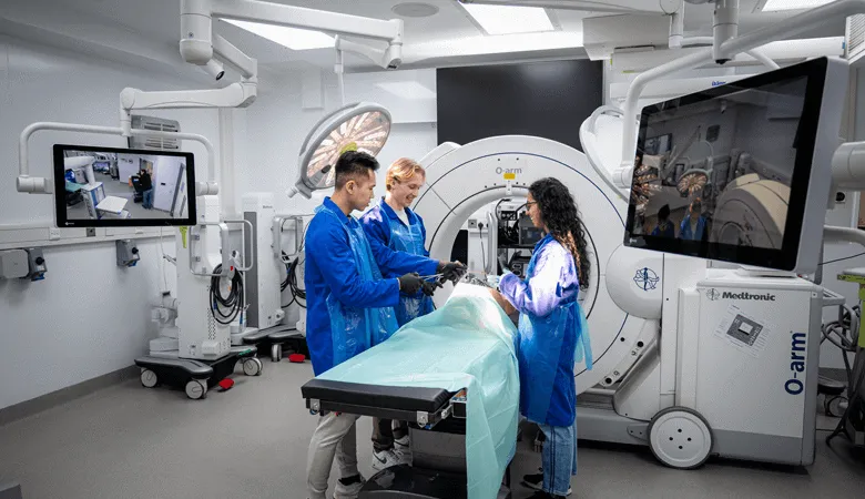
The micro-CT scanner uses X-rays to obtain high-resolution 3D images of small specimens from 3mm to 45mm in diameter and up to 450mm in length. Images can be captured of both hard tissue (e.g. bones, teeth) and soft tissue (with the aid of a suitable stain). Voxel size can be adjusted from 1 to 45 microns.
Access: Contact the facility manager for more information on scanning samples.
Training: Training is provided by the facility technician. Users will need to complete the 'Basic Ionising Radiation Safety' module online prior to access.
Location: Guy's Hospital, 27th floor, Tower Wing, Great Maze Pond, London SE1 1UL | Guy’s Campus
Contact: Facility Team: techteam-ccrb@kcl.ac.uk or Academic Lead: Abigail Tucker
The Zeiss LSM980 is equipped with 6 lasers (405/445/488/514/561/639nm) and 4 objectives (10x/20x/40xW/63xO), as well as an Airyscan.
Access: The facility is available to King’s researchers. Contact the Facility Manager for access.
Training: Two to three training sessions are provided by the Facility Manager, including time to optimise individual samples, before a user can access the facility independently.
Location: Guy's Hospital, 27th floor, Tower Wing, Great Maze Pond, London SE1 1UL | Guy’s Campus
Contact: techteam-ccrb@kcl.ac.uk
The CDN has two confocal microscopes, a Lightsheet (LSFM) and a Slide Scanner, available to users.
Equipment details:
Access: Contact the facility manager
Training: Training sessions are provided to operate equipment independently
Location: Centre for Developmental Neurobiology, New Hunt’s House, Great Maze Pond, London SE1 1UL | Guy’s Campus
Contact: Facility Manager: Anneliese Jarman
A centre of expertise for cell characterisation using high throughput imaging and high content analysis of cellular models. The stem cell hotel provides guidance, expert advice and bespoke services to users, including assay development and assistance in overcoming technical challenges. It advises researchers in the best practice of sample preparation for imaging, imaging expertise, analysis pipeline design and quantification of the results. Introductory training is provided to all new users and bespoke training for complex cellular models is available.
Access: To access the booking system, create an online account by completing the registration form.
Training: To qualify as an independent user, individual training can be arranged based on project requirements.
Location: 28th floor, Centre for Gene Therapy and Regenerative Medicine, Tower Wing, Guy’s Hospital, London SE19RT| Guy’s Campus
Contact: Complete the online enquires form or email the facility at info-sch@kcl.ac.uk
The Centre for Ultrastructural Imaging (CUI) is the central electron microscopy core research facility at King’s, offering electron microscope (EM) services for internal and external collaborators from academic, commercial and industrial fields. Using advanced EM instruments, we perform and support various workflows, including room temperature preparation, through to full cryo-preparation and imaging, multi-scale correlative and volume imaging.
Access: Contact the Centre.
Training: Individual training for users to operate microscopes and prepare specimens is provided and the CUI hold regular seminars and workshops.
Location: New Hunt’s House, Great Maze Pond, London SE1 1UL | Guy’s Campus
Contact: cui@kcl.ac.uk
The ExoviewR200 facility allow users the sensitive detection & characterisation of extracellular vesicles and viruses, including exosomes and lentiviruses. The fully automated platform provides comprehensive measurements for particles; size analysis, concentration, phenotype, and biomarker colocalization.
Access: Contact facility manager for enquiries
Training: The facility provides complete training by one of their experts
Location: James Black Centre, 125 Coldharbour Lane, London, SE5 9NU | 8AF | Denmark Hill Campus
Contact: Facility Manager: Sadia Ahmad
The HTS facility is equipped with state-of-the-art instrumentation to perform high-throughput and high-content functional genomics screenings using a range of libraries. The platform can accommodate cell-based screenings using diverse cell models to address a wide range of biological questions.
Access: Contact the facility manager to discuss project requirements.
Location: The James Black Building, 125 Coldharbour Lane, London SE5 9NU | Denmark Hill Campus
Contact: Facility Manager Miguel Mano
The imaging clinical research facility includes nine advanced MRI systems (0.064T, 0.55T. 1.5T, 3T, 7T) that are used to deliver clinical services to more than 5,000 patients per year, as well as a diverse range of imaging research projects. Strengths are in cardiovascular, perinatal and ultra-high-field sub-specialties.
Access: Researchers may gain access to the scan suites that make up the Imaging Clinical Research Facility by requesting approval for an imaging project and completing a set of mandatory requirements, including MRI safety training to enable independent or supervised access.
Training: KHP passport and completion of GSTT statutory and mandatory training modules if access to clinical data / patients + completion of KCL workrite modules, MRI safety training and evacuation training + GCP training
Location: Lambeth Wing, St Thomas Hospital, Westminster Bridge Road, London SE1 7EH
Contact: Facility Manager Ramesh Valapil
King's College London & Guy's and St Thomas' PET Centre provides PET services for referred patients at a local and national level and performs clinically related research. The Centre hosts three clinical PET-CTs and one simultaneous PET-MR, cyclotron and radiochemistry facilities, and associated equipment for state-of-the art imaging research.
Access: Visit the PET Centre webpage for further details.
Location: Lambeth Wing, St Thomas Hospital, Westminster Bridge Road, London SE1 7EH
Contact: gst-tr.PETCentre@nhs.net
The Microscopy Innovation Centre is a core research facility that collaborates with academic and industrial partners to develop and house advanced microscopy instrumentation and analysis tools, offering cutting-edge microscopes to support advanced imaging.
Access: The MIC access policy includes details on access and eligibility.
Training: Individual training sessions are provided for all microscopes.
Location: Hodgkin Building, Great Maze Pond, London SE1 1UL | Guy’s Campus
Contact: Facility Manager Andreas Bodén, or Deputy Manager Dylan Herzog
This facility enables users to undertake parallel analysis of the two major energy pathways in the cells mitochondrial respiration and glycolysis, in real time, quantifying oxidative phosphorylation, fatty acid oxidation, extracellular acidification, glycolysis, substrate utilisation evaluation and other key bioenergetic measurements in live cells. The instruments available are:
Visit Agilent for help with designing your experiments.
Access: Contact the Facility Manager to discuss access.
Training: Training is required for access to the facility and the equipment booking system. To book onto the next training sessions, email Aakruti Kaikini. Hands on training takes place over 2 days and is provided by an Agilent expert. This is followed by a data handling training session.
Location: Hodgkin Building, Great Maze Pond, London SE1 1UL | Guy’s Campus
Contact: Facility Manager: Dr Hannah Rosa
Providing facilities for both basic neuroscience and more applied clinical research studies, incorporating structural, functional, pharmacological MRI techniques. A dedicated team support the development of fMRI paradigms and associated ancillary equipment, and we can provide access to a range of clinical and non-clinical spaces for use during scanning sessions.
Access: Contact the facility for enquiries and costings for grant applications.
Training: Individual consultations to discuss technical and logistical requirements and pilot scanning session provided.
Location: Centre for Neuroimaging Sciences, DeCrespigny Park, London, SE5 8AF | Denmark Hill Campus
Contact: scanningcostings@kcl.ac.uk
The Nikon Imaging Centre, (NIC@King's) is a core research facility for light microscopy developed in partnership between King’s College London and Nikon Instruments UK. Introductory training and ongoing support is provided and tailored to user needs.
Access: New users can create an account and book training via the NIC@King's webpage, or to arrange a consultation, contact the facility manager.
Training: 2/3 tailored training sessions are provided to support project initiation and and training to operate equipment independently.
Location: Hodgkin Building, Great Maze Pond, London SE1 1UL | Guy’s Campus
Contact: Facility Manager, Chantal Hubens
Biomarker Research and Imaging for Neuroscience (The BRAIN Centre) is a world leading facility for preclinical in vivo and ex vivo neuro-imaging of experimental animal models of neurological and psychiatric disorders. While the main focus is on magnetic resonance (MR) – structural & functional imaging and spectroscopy, the centre also provides assistance with small animal brain PET scanning as well as variety of post-mortem imaging and in vivo techniques such as microdialysis, electroencephalography, behavioural testing, histology and autoradiography.
Access: Contact the facility for enquiries and costings for grant applications.
Training: Full training is provided, or assistance with imaging experiments is also possible for untrained or inexperienced staff or students. Contact the facility for more information.
Location: The BRAIN Centre, Neuroimaging, James Black Centre, 125 Coldharbour Lane, London SE5 9NU | Denmark Hill Campus
Contact: Facility Manager: Dr Diana Cash or email the brain@kcl.ac.uk
The London Metallomics Facility (LMF) is a core research facility that provides analytical tools to understand the critical roles that metals play in biology with the ability to quantify and spatially map elemental isotopes in a wide range of challenging matrices using a combination of ICP-MS, LA-ICP-MS/MS, HPLC, and TXRF for solid and solution based samples.
Access: Once users have created an account, they can book services and equipment via the online booking portal.
Training: Regular symposiums available showcasing recent developments. Check the LMF webpage for details.
Location: Franklin Wilkins Building, Stamford St, London SE1 9NQ | Waterloo Campus
Contact: lmf@kcl.ac.uk
The Wohl Cellular Imaging Centre (WCIC) provides cutting edge light microscopy equipment, including confocal, multiphoton, light sheet and super-resolution microscopes, as well as tailored training for microscopical imaging and image analysis software and workstations.
Access: A facility induction is provided.
Training: Bespoke microscope acquisition and analysis training, plus a wide range of online learning resources, included courses available on KEATS for King's students and staff.
Location: Maurice Wohl Clinical Neuroscience Institute, 8 Cutcombe Road, Camberwell, London, SE5 9RT
Contact: Facility Manager: George Chennell
X-ray crystallography analyses the x-ray diffraction patterns and quality of crystallisation to determine the atomic and molecular structure of protein in crystals.
Access: Email Risa Mori for facility enquiries.
Training: Individual training is provided for use of equipment.
Location: New Hunt’s House, Great Maze Pond, London, London SE1 1UL | Guy’s Campus
Contact: Facility Manager: Professor Brian Sutton
Cutting-edge facilities, technical services and technology platforms.